. 2020 Jan 20;12(1):15.
doi: 10.1186/s13148-020-0807-x.

A DNA methylation signature to improve survival prediction of gastric cancer

Affiliations

A DNA methylation signature to improve survival prediction of gastric cancer

Yaojun Peng et al. Clin Epigenetics. .

Abstract

Background: The current Union International Committee on Cancer or the American Joint Committee on Cancer TNM stage system has shown valuable but insufficient estimation for subsets of gastric cancer and prediction for prognosis patients. Thus, there is an urgent need to identify diagnostic, prognostic, and predictive biomarkers to improve patients' outcomes. Our aim was to perform an integrative analysis on publicly available datasets to identify epigenetic changes that may play key role in the initiation and progression of gastric cancer, based on which we set to develop a DNA methylation signature to improve survival prediction of gastric cancer.

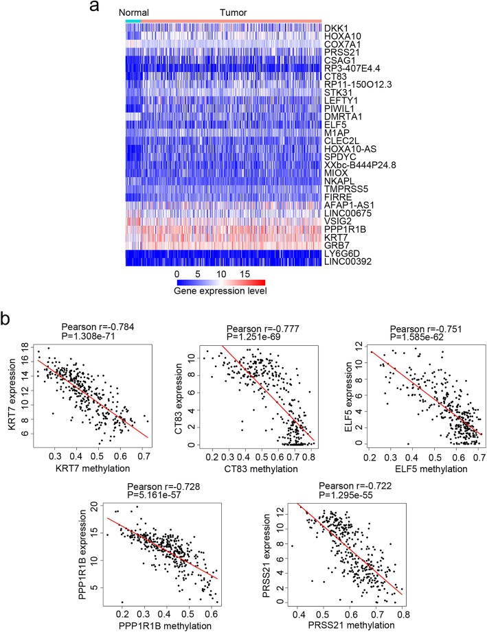
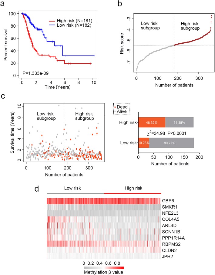
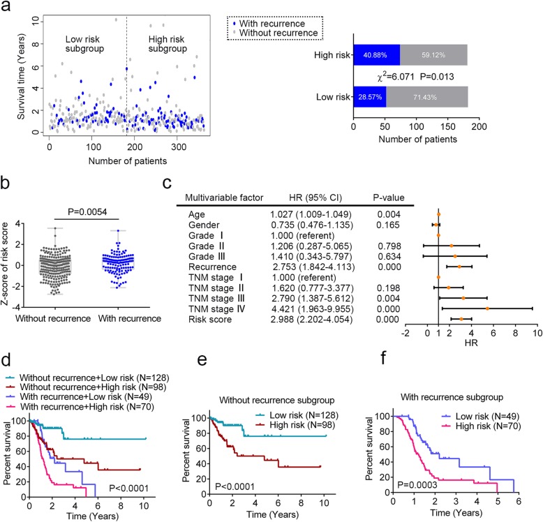
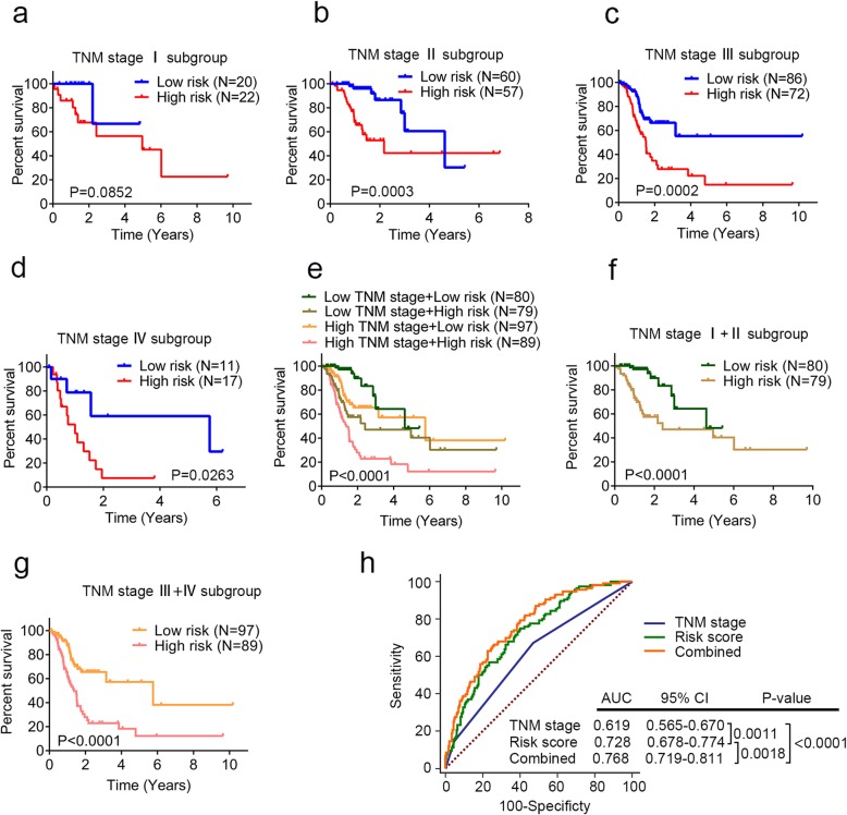
Results: A total of 340 methylation-related differentially expression genes (mrDEGs) were screened in gastric cancer patients from The Cancer Genome Atlas (TCGA) project. Pathway enrichment analysis revealed that they were involved in the biological process related to initiation and progression of gastric cancer. Based on the mrDEGs identified, we developed a DNA methylation signature consisting of ten gene members (SCNN1B, NFE2L3, CLDN2, RBPMS2, JPH2, GBP6, COL4A5, SMKR1, PPP1R14A, and ARL4D) according to their methylation β value. This innovative DNA methylation signature was associated with cancer recurrence, while it showed independence of cancer recurrence and TNM stage for survival prediction. Combination of this DNA methylation signature and TNM stage improved overall survival prediction in the receiver operating characteristic analysis. We also verified that two individual genes (PPP1R14A and SCNN1B) of the identified prognostic signature were regulated by promoter region methylation in a panel of gastric cell lines.

Conclusions: This study presents a powerful DNA methylation signature by performing analyses integrating multi-source data including transcriptome, methylome, and clinical outcome of gastric cancer patients from TCGA. The identified DNA methylation signature may be used to refine the current prognostic model and facilitate further stratification of patients in the future clinical trials. Further experimental studies are warranted to unveil the regulatory mechanism and functional role of all the individual genes of the DNA methylation signature. Also, clinical investigations in large GC patient cohorts are greatly needed to validate our findings.

Keywords: DNA methylation; Epigenetics; Gastric cancer; Integrative analysis; Prognosis; TCGA.


Conflict of interest statement

The authors declare that they have no competing interests.

Figures

Fig. 1
Fig. 1
Determining methylation related differentially expressed genes (mrDEGs) in gastric cancer (GC). a The expression profile of the most significant 30 mrDEGs between normal (N = 32) and GC samples (N = 375). b The association between gene expression and DNA methylation of the top 5 mrDEGs in GC samples (N = 338) whose expression and DNA methylation data were both available
Fig. 2
Fig. 2
Pathway enrichment analysis of the upregulated and downregulated methylation related differentially expressed genes (mrDEGs) in gastric cancer. a The pathway enrichment results of the upregulated mrDEGs. b The pathway enrichment results of the downregulated mrDEGs. Each node represents one enriched term. Node size is proportional to the total number of genes within each gene set. Proportion of shared genes between gene sets is represented as the thickness of the line between nodes
Fig. 3
Fig. 3
The DNA methylation signature for overall survival (OS) prediction of gastric cancer (GC) patients. a Kaplan-Meier estimate of the OS using the identified DNA methylation signature. GC patients were divided into low-risk (N = 181) or high-risk (N = 182) subgroup based on the median of risk score. The difference between the two curves was determined by the two-side log-rank test. b The distribution of risk score derived from the DNA methylation signature. c The distribution of GC patients’ survival status. The difference between the low-risk and high-risk subgroup was determined by chi-square test. d The methylation β value profile of the identified DNA methylation signature in the high-risk and low-risk subgroups
Fig. 4
Fig. 4
The DNA methylation signature is associated with cancer recurrence. a The distribution of cancer recurrence status in the high-risk and low-risk subgroup. The difference between the two subgroups was determined by chi-square test. b Box plot of risk score of patients with or without cancer recurrence. T test was used to determine the significance of the comparison. c The multivariate Cox regression analysis performed on 345 gastric cancer (GC) patients that contained age, gender, tumor grade, cancer recurrence, TNM stage, and risk score as covariates. Risk score and age were evaluated as continuous variables, and gender, tumor grade, cancer recurrence, and TNM stage were evaluated as category variables. Orange solid dots represent the hazard ratio (HR) of death and open-ended horizontal lines represent the 95% confidence intervals (CIs). All P values were calculated using Cox proportional hazards analysis. d Kaplan-Meier estimate of the overall survival of the entire set GC patients (N = 345) using the DNA methylation signature. GC patients were stratified by cancer recurrence status and the high-risk or low-risk subgroup of patients was determined on the basis of the median risk score. e Kaplan-Meier curves for GC patients without cancer recurrence (N = 226). f Kaplan-Meier curves for patients with cancer recurrence (N = 119). The differences between the survival curves were determined by the two-sided log-rank test
Fig. 5
Fig. 5
Prognostic value of the DNA methylation signature is independent of TNM stage. a Kaplan-Meier curves for patients with TNM stage I (N = 42). Gastric cancer (GC) patients were stratified by TNM stage (I, II, III, and IV) and the high-risk or low-risk subgroup of patients was determined on the basis of the median risk score. b Kaplan-Meier curves for patients with TNM stage II (N = 117). c Kaplan-Meier curves for patients with TNM stage III (N = 158). d Kaplan-Meier curves for patients with TNM stage IV (N = 28). e Kaplan-Meier estimate of the overall survival (OS) of the entire set GC patients (N = 345) using the DNA methylation signature. GC patients were stratified by TNM stage (I + II and III + IV). f Kaplan-Meier curves for patients with low TNM stage (stage I + II, N = 159). g Kaplan-Meier curves for patients with high TNM stage (stage III + IV, N = 186). The differences between the survival curves were determined by the two-sided log-rank test. h Receiver operating characteristic (ROC) analysis of the sensitivity and specificity of OS prediction by the DNA methylation signature risk score, TNM stage, and combination of the two factors. P values were obtained from the comparisons of the area under the ROC (AUROC) of DNA methylation signature risk score versus those of TNM stage and DNA methylation risk score combined with TNM stage
Fig. 6
Fig. 6
The expression of PPP1R14A and SCNN1B, two individual genes of the identified prognostic signature, are regulated by promoter region methylation. a Expression levels of PPP1R14A and SCNN1B without (−) or with (+) 5-aza treatment were analyzed by qPCR in eight gastric cell lines (GES1, NUGC3, SNU5, SNU16, NCI-N87, AGS, MGC803, and BGC823). b Schematic diagrams of CpG islands in the promoter region of PPP1R14A and SCNN1B. MF methylation forward primer, MR methylation reverse primer, UF unmethylation forward primer, UR unmethylation reverse primer, BSSQ-F bisulfite sequencing forward primer, BSSQ-R bisulfite sequencing reverse primer. c Methylation status of PPP1R14A and SCNN1B was detected by methylation specific PCR (MSP) in gastric cell lines. IVD in vitro methylated DNA, NL normal lymphocyte DNA, M methylated alleles, U unmethylated alleles. d Bisulfide sequencing (BSSQ) of PPP1R14A was performed in GES1 and AGS cell lines. For SCNN1B, GES1 and SNU16 cell lines were analyzed. Red solid dots represent methylated CpG sites, and green solid dots denote unmethylated CpG sites. The horizontal black bar demarcates the primers of MSP, which are included in the region of BSSQ
Fig. 7
Fig. 7
Flowchart showing steps involved in identification of the prognostic DNA methylation signature in gastric cancer

References

    1. Bray F, Ferlay J, Soerjomataram I, Siegel RL, Torre LA, Jemal A. Global cancer statistics 2018: GLOBOCAN estimates of incidence and mortality worldwide for 36 cancers in 185 countries. CA Cancer J Clin. 2018;68(6):394–424. doi: 10.3322/caac.21492. - DOI - PubMed
    1. Van Cutsem E, Sagaert X, Topal B, Haustermans K, Prenen H. Gastric cancer. Lancet (London, England) 2016;388(10060):2654–2664. doi: 10.1016/S0140-6736(16)30354-3. - DOI - PubMed
    1. Lott PC, Carvajal-Carmona LG. Resolving gastric cancer aetiology: an update in genetic predisposition. The lancet Gastroenterology & hepatology. 2018;3(12):874–883. doi: 10.1016/S2468-1253(18)30237-1. - DOI - PMC - PubMed
    1. Tan P, Yeoh KG. Genetics and molecular pathogenesis of Gastric adenocarcinoma. Gastroenterology. 2015;149(5):1153–62.e3. doi: 10.1053/j.gastro.2015.05.059. - DOI - PubMed
    1. Dikken JL, van de Velde CJ, Coit DG, Shah MA, Verheij M, Cats A. Treatment of resectable gastric cancer. Therapeutic advances in gastroenterology. 2012;5(1):49–69. doi: 10.1177/1756283X11410771. - DOI - PMC - PubMed

Publication types

MeSH terms

Substances