Review
. 2020:136:3-32.
doi: 10.1016/bs.ctdb.2019.11.004. Epub 2019 Dec 12.

Setting up for gastrulation: D. melanogaster

Affiliations
Review

Setting up for gastrulation: D. melanogaster

Angelike Stathopoulos et al. Curr Top Dev Biol. 2020.

Abstract

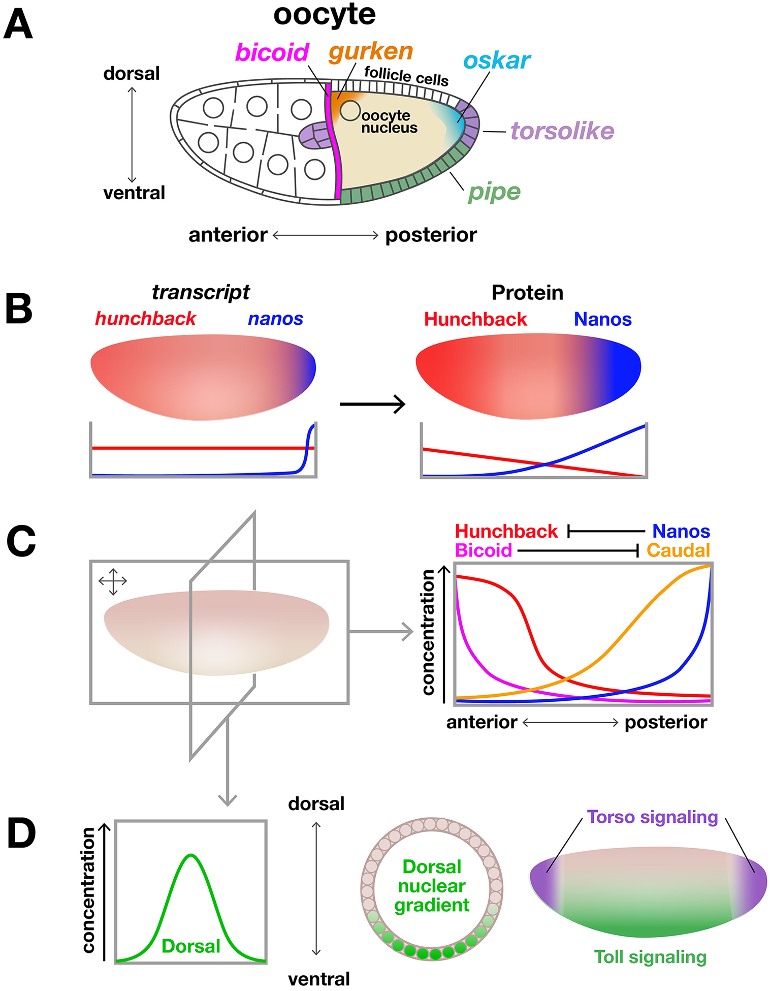
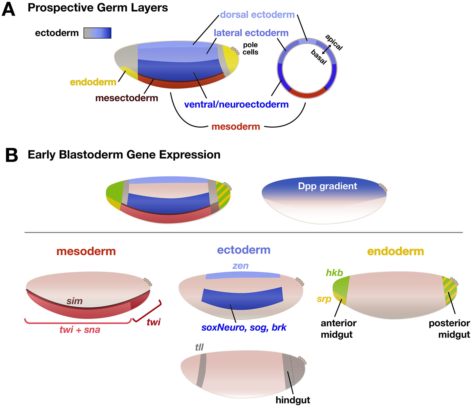
Drosophila melanogaster embryos develop initially as a syncytium of totipotent nuclei and subsequently, once cellularized, undergo morphogenetic movements associated with gastrulation to generate the three somatic germ layers of the embryo: mesoderm, ectoderm, and endoderm. In this chapter, we focus on the first phase of gastrulation in Drosophila involving patterning of early embryos when cells differentiate their gene expression programs. This patterning process requires coordination of multiple developmental processes including genome reprogramming at the maternal-to-zygotic transition, combinatorial action of transcription factors to support distinct gene expression, and dynamic feedback between this genetic patterning by transcription factors and changes in cell morphology. We discuss the gene regulatory programs acting during patterning to specify the three germ layers, which involve the regulation of spatiotemporal gene expression coupled to physical tissue morphogenesis.

Keywords: Anterior-posterior patterning; Dorsal-ventral patterning; Drosophila melanogaster; Ectoderm; Embryonic development; Endoderm; Gastrulation; Germ-band elongation; Maternal-to-zygotic transition; Mesoderm; Morphogen gradients; Syncytium.


Figures

Figure 1 -
Figure 1 -. Establishment of embryonic polarity
A) Key maternal transcripts are specifically localized within the oocyte by RNA-binding proteins establishing basic polarity that is transferred to the zygote. B) Oskar-dependent posterior localization of nanos transcript establishes an anterior-to-posterior gradient of Hunchback protein through translational repression of uniformly-distributed hunchback mRNA. C) Schematic of distribution of four key patterning factors along the A-P axis of the pre-blastoderm embryo. D) D-V biased Toll signaling resulting from Pipe localization in the ventral oocyte produces a nuclear gradient of the transcription factor Dorsal. tsl transcript deposited to the anterior and posterior of the oocyte ultimately activates Torso signaling in corresponding terminal domains in the early embryo. Note: all follicle and embryo diagrams are oriented with anterior left and dorsal up unless otherwise specified.
Figure 2 -
Figure 2 -. Maternal to Zygotic Transition
A) Early zygotic development entails 13 rapid nuclear divisions within a common cytoplasm to produce a syncytium. Nuclei migrate to the embryo periphery and then cellularize forming a blastoderm of ~6000 cells surrounding a central yolk. In order to transition from control of development by maternal RNA and proteins to regulation driven by zytotic products, maternal factors are systematically cleared from the embryo in two phases. B) Maternal transcripts deposited into the zygote affect early development and the onset of Zygotic Genome Activation (ZGA) by their precise localization within the embryo, stabilization and translation of RNA into factors that regulate key phenomena: the repression and degradation of maternal factors, regulation of nuclear divisions as early blastoderm development proceeds, and activation of zygotic gene expression. C) Models for timing of ZGA. Top: “Maternal Clock” model - time required for buildup of key activators determines onset of ZGA. Middle: Chromatin Accessibility model - as nuclei divide, the concentration of soluble histones declines. Combined with other mechanisms, including activity of pioneer factors, chromatin accessibility (particularly at enhancers) increases and zygotic genes begin to be expressed. Bottom: Repressor Titration - as the ratio of nuclei to cytoplasm increases during early divisions, concentration of maternal repressors is diluted within individual nuclei; once below repression threshold, zygotic targets of repression begin to be expressed. Note: Models shown are conceptual, exact timing of ZGA and levels of relevant factors likely differ depending on context. Adapted from Hamm & Harrison, 2018.
Figure 3 -
Figure 3 -. Gene expression patterns establish prospective germ layers
A) Broad patterns of gene expression subdivide the embryo into domains that prefigure the germ layers. B) Ventral cells specified by expression of twist and snail and delineated by expression of sim will develop into the mesoderm. Domains along the trunk dorsal to sim comprise the future ectoderm. A dorsal gradient of Dpp signaling contributes to further subdivision of the ectodermal primordia into ventral/neuroectoderm and non-neuroectoderm (the lateral ectoderm and dorsal ectoderm/amnioserosa). Anterior and posterior ectodermal domains marked by tll will form the fore- and hindgut, respectively. Domains at the embryo termini marked by the expression of hkb, tll, and srp will form the endoderm.
Figure 4 -
Figure 4 -. Gene regulatory interactions prepare cells for diverse cell movements at gastrulation
(A) Mesoderm Invagination: Expression of Twist and Snail lead to apical constriction which generates a ventral furrow (B) Endoderm Invagination: The anterior and posterior endoderm are specified by distinct mechanisms and invaginate separately. They eventually merge to form the gut. (C) Germ-band Extension: GBE is largely driven by cell intercalation and cell shape changes that are a passive response to extrinsic tensile forces. Cells undergo convergent extension, shape changes and intercalation which leads to the overall lengthening of the ectoderm along the A-P axis, orthogonal to Toll gene expression (not shown). (D) Cephalic Furrow Formation: eve expression leads to myosin-independent cell shortening. Actomyosin-independent dorsal fold formation occurs at stripes of runt expression and results from basal junctional repositioning. Note: For simplicity, only the relevant stripes of eve and runt expression are shown. Adapted from Gilmour, Rembold, & Leptin, 2017.

References

    1. Ajduk A, & Zernicka-Goetz M (2016). Polarity and cell division orientation in the cleavage embryo: from worm to human. Molecular Human Reproduction, 22(10), 691–703. - PMC - PubMed
    1. Alberga A, Boulay JL, Kempe E, Dennefeld C, & Haenlin M (1991). The snail gene required for mesoderm formation in Drosophila is expressed dynamically in derivatives of all three germ layers. Development, 111(4), 983–992. - PubMed
    1. Ali-Murthy Z, & Kornberg TB (2016). Bicoid gradient formation and function in the Drosophila pre-syncytial blastoderm. eLife, 5 10.7554/eLife.13222 - DOI - PMC - PubMed
    1. Ali-Murthy Z, Lott SE, Eisen MB, & Kornberg TB (2013). An essential role for zygotic expression in the pre-cellular Drosophila embryo. PLoS Genetics, 9(4), e1003428. - PMC - PubMed
    1. Amarnath S, Stevens LM, & Stein DS (2017). Reconstitution of Torso signaling in cultured cells suggests a role for both Trunk and Torso-like in receptor activation. Development, 144(4), 677–686. - PMC - PubMed

Publication types

LinkOut - more resources