Review
. 2020 Mar;26(3):285-295.
doi: 10.1016/j.molmed.2019.11.008. Epub 2020 Jan 18.

Glymphatic System Impairment in Alzheimer's Disease and Idiopathic Normal Pressure Hydrocephalus

Affiliations
Review

Glymphatic System Impairment in Alzheimer's Disease and Idiopathic Normal Pressure Hydrocephalus

Benjamin C Reeves et al. Trends Mol Med. 2020 Mar.

Abstract

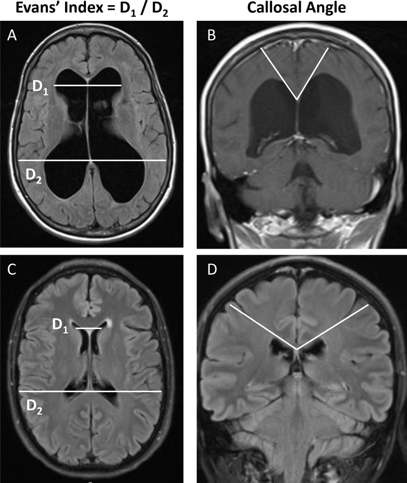
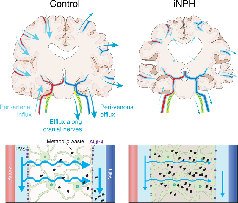
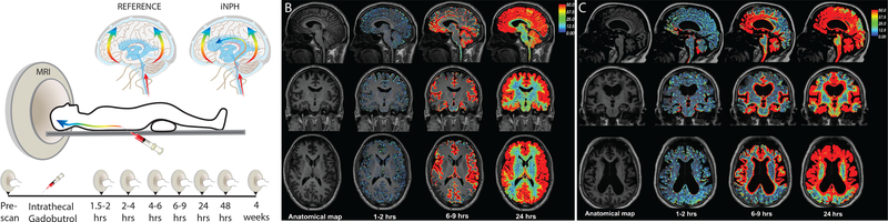
Approximately 10% of dementia patients have idiopathic normal pressure hydrocephalus (iNPH), an expansion of the cerebrospinal fluid (CSF)-filled brain ventricles. iNPH and Alzheimer's disease (AD) both exhibit sleep disturbances, build-up of brain metabolic wastes and amyloid-β (Aβ) plaques, perivascular reactive astrogliosis, and mislocalization of astrocyte aquaporin-4 (AQP4). The glia-lymphatic (glymphatic) system facilitates brain fluid clearance and waste removal during sleep via glia-supported perivascular channels. Human studies have implicated impaired glymphatic function in both AD and iNPH. Continued investigation into the role of glymphatic system biology in AD and iNPH models could lead to new strategies to improve brain health by restoring homeostatic brain metabolism and CSF dynamics.

Keywords: Alzheimer’s disease; aging; dementia; glial-lymphatic; glymphatic; hydrocephalus; idiopathic normal pressure hydrocephalus.


Figures

Figure 1:
Figure 1:. T1-Weighted MRIs showing cardinal diagnostic features of iNPH.
Axial and coronal brain MRIs of an individual diagnosed with iNPH (A, B) compared to brain MRIs of an age and sex-matched control (C, D). Note the key diagnostic features of iNPH: ventriculomegaly determined by an increased Evan’s index and a steeper callosal angle.
Figure 2:
Figure 2:. Schematic of potential glymphatic dysfunction in iNPH.
Top Panel – Diagram showing the periarterial CSF influx routes and proposed perivenous and perineural cerebrospinal (CSF) efflux routes of the glymphatic system in a control (left) and iNPH (right) cortical brain section. The less prominent arrows in the iNPH brain denote reduced CSF flux along the perivascular and perineural pathways demonstrated by Ringstad and Per Eide in their human MRI studies. The arrows in the iNPH brain also demonstrate transependymal flow of CSF from the lateral ventricles into the brain parenchyma. Bottom Panel – Schematic demonstrating glymphatic system function in a control (left) brain and iNPH brain (right). Reduced arrow sizes in the iNPH schematic indicate attenuated CSF periarterial inflow, CSF-interstitial fluid (ISF) exchange, and perivenous efflux of CSF mediated by the depolarization of AQP4 that has been observed in human iNPH patients. This reduction in the exchange of CSF and ISF causes increased concentrations of neuronal metabolic waste products in the brain interstitial space. Note: The periarterial influx routes shown on the left side of the control brain and the perivenous efflux routes shown on the right of the control brain are for illustrative purposes only and do not imply that fluid enters on one side of the brain and leaves on the opposite side.
Figure 3:
Figure 3:. Impaired glymphatic CSF transport in hydrocephalus.
(A) Repeated contrast-enhanced magnetic resonance imaging (MRI) after intrathecal delivery of gadobutrol. The contrast agent distributed into all parts of the brain over 24 hrs in 9 iNPH and 8 reference patients. The diagrams illustrate gadobutrol dispersal in a centripetal pattern along the large cerebral arteries in both patient groups, but enrichment in periventricular white matter only in idiopathic normal pressure hydrocephalus (iNPH) patients, presumably reflecting ventricular CSF reflux. (B) Repeated scans of a reference patient display brain-wide distribution of the contrast agent 24 hrs after intrathecal delivery. (C) An iNPH patient also exhibits brain-wide contrast enrichment at 24 hrs. Enhanced gadobutrol uptake was evident in iNPH patients in periventricular white matter. Gadobutrol clearance was significantly delayed in iNPH compared with reference patients at 24 hrs, but all 17 subjects cleared the contrast agent at 4 weeks (data not shown). Modified from Ringstad et al., 2018, with permission.

Comment in

Similar articles

Cited by

References

    1. International D and Patterson C (2018) World Alzheimer Report 2018 - The state of the art of dementia research: New frontiers. Alzheimer’s Dis. Int DOI: 10.1103/PhysRevLett.78.4414 - DOI
    1. Nassar BR and Lippa CF (2016) Idiopathic Normal Pressure Hydrocephalus: A Review for General Practitioners. Gerontol. Geriatr. Med 2, 2333721416643702. - PMC - PubMed
    1. Kiefer M and Unterberg A (2012) The Differential Diagnosis and Treatment of Normal-Pressure Hydrocephalus. Dtsch. Arztebl. Int 109, 15–26 - PMC - PubMed
    1. Wallenstein MB and McKhann GM (2010) Salomón Hakim and the discovery of normal-pressure hydrocephalus. Neurosurgery 67, 155–159 - PubMed
    1. Martín-Láez R et al. (2016) Incidence of Idiopathic Normal-Pressure Hydrocephalus in Northern Spain. World Neurosurg. DOI: 10.1016/j.wneu.2015.10.069 - DOI - PubMed