. 2020 Mar 19;77(6):1265-1278.e7.
doi: 10.1016/j.molcel.2019.12.019. Epub 2020 Jan 17.

A Dimeric Structural Scaffold for PRC2-PCL Targeting to CpG Island Chromatin

Affiliations

A Dimeric Structural Scaffold for PRC2-PCL Targeting to CpG Island Chromatin

Siming Chen et al. Mol Cell. .

Abstract

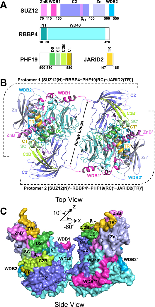
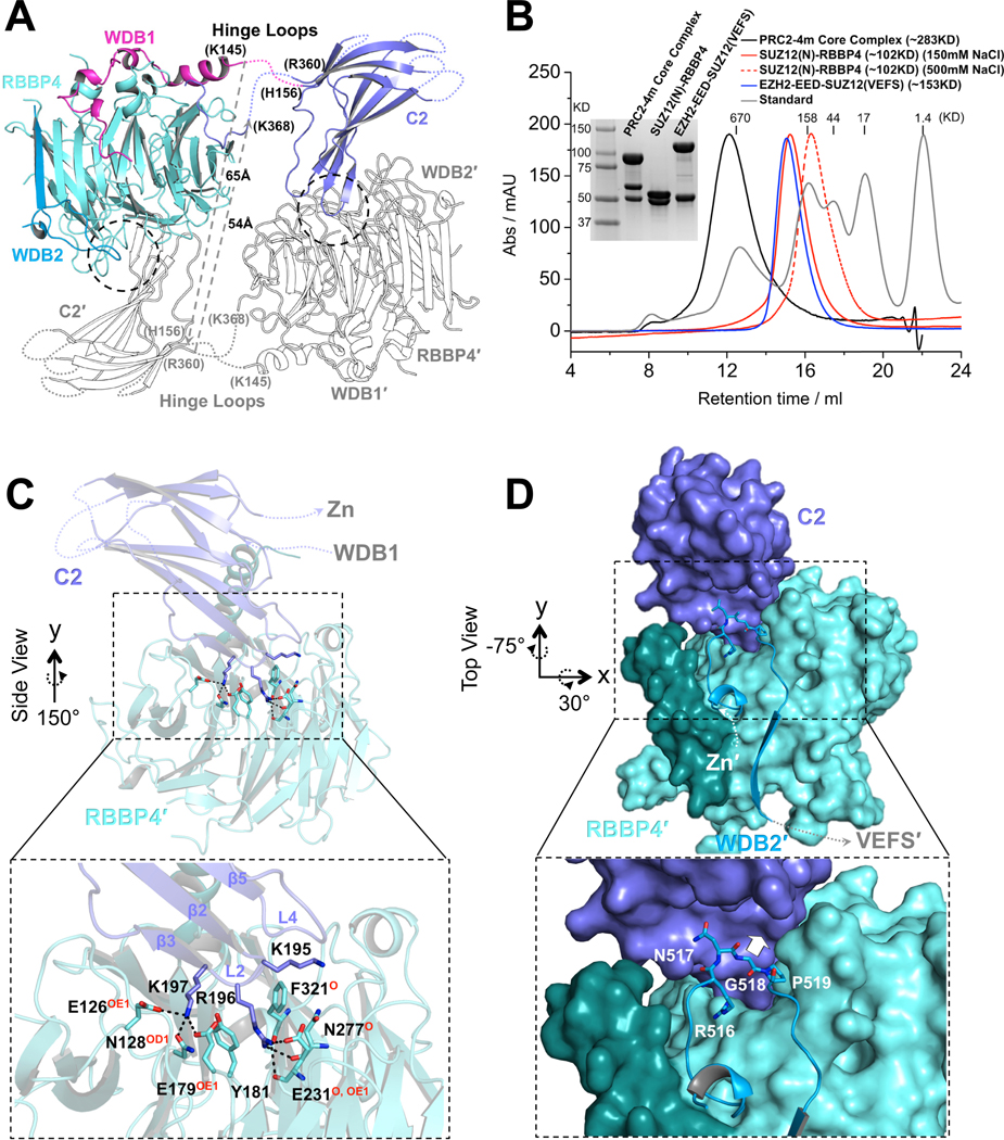
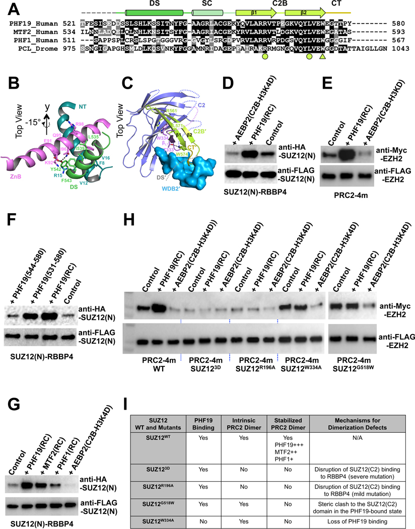
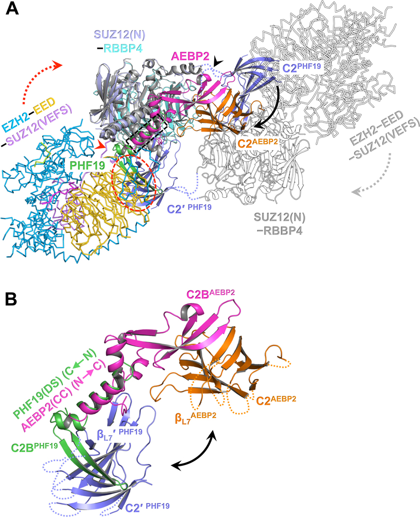
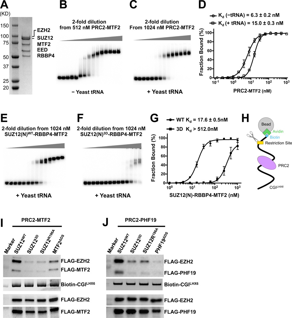
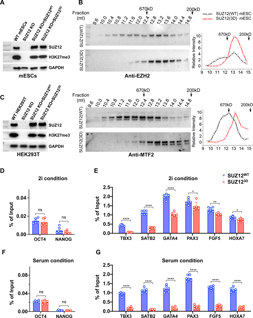
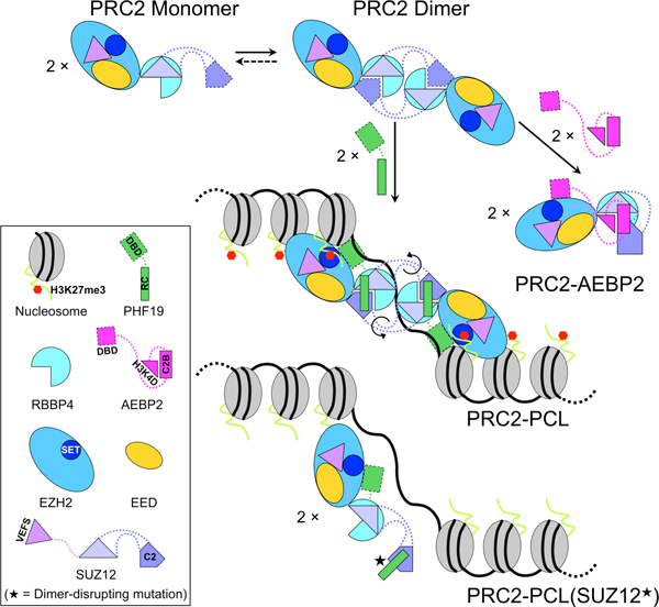
Diverse accessory subunits are involved in the recruitment of polycomb repressive complex 2 (PRC2) to CpG island (CGI) chromatin. Here we report the crystal structure of a SUZ12-RBBP4 complex bound to fragments of the accessory subunits PHF19 and JARID2. Unexpectedly, this complex adopts a dimeric structural architecture, accounting for PRC2 self-association that has long been implicated. The intrinsic PRC2 dimer is formed via domain swapping involving RBBP4 and the unique C2 domain of SUZ12. MTF2 and PHF19 associate with PRC2 at around the dimer interface and stabilize the dimer. Conversely, AEBP2 binding results in a drastic movement of the C2 domain, disrupting the intrinsic PRC2 dimer. PRC2 dimerization enhances CGI DNA binding by PCLs in pairs in vitro, reminiscent of the widespread phenomenon of transcription factor dimerization in active transcription. Loss of PRC2 dimerization impairs histone H3K27 trimethylation (H3K27me3) on chromatin at developmental gene loci in mouse embryonic stem cells.

Keywords: CpG island (CGI); chromatin; crystal structure; epigenetics; gene silencing; histone methylation; polycomb repressive complex 2 (PRC2); polycomb-like (PCL); protein dimerization; structural biology.


Conflict of interest statement

Declaration of Interests The authors declare no competing interests.

Figures

Fig. 1
Fig. 1. Overall structure of the SUZ12(N)-RBBP4-PHF19(RC)-JARID2(TR) heterotetrameric complex in the dimeric state
(A) Domain structures of the proteins in the crystal. ZnB, Zinc-finger Binding; WDB1, WD40-Binding 1; Zn, Zinc-finger; WDB2, WD40-Binding 2; NT, N-Terminal; DS, Dimer Stabilization; SC, Short Connecting; C2B, C2-Binding; CT, C-Terminal; TR, Transrepression. C2 and βL7 are not abbreviated names. (B) Cartoon representation of the dimeric complex in Top View. Disordered protein loops are displayed as dotted lines. Hinge loops that connect to the C2 domains are indicated by black arrows. The second protomer of the dimer is labeled with a prime symbol. (C) Surface representation of the dimeric complex in Side View. The rotational matrix that relates the Top View and Side View is indicated. See also Fig. S1.
Fig. 2
Fig. 2. Dimerization of the PRC2 core complex
(A) Structure of one protomer of the PRC2 core complex in cartoon representation. Only SUZ12(N) and RBBP4 are shown. Domains within only one protomer are colored. The SUZ12(ZnB) and SUZ12(Zn) domains are removed for clarity. SUZ12 residues at the junction of the disordered hinge loops are labeled. The minimal lengths of a pair of hypothetical hinge loops between the WDB1 domain from one protomer and the C2 domain from the other protomer that would exist in a closed complex are indicated by dotted gray lines. (B) SEC elution profiles of the four-member PRC2 core complex (PRC2–4m), SUZ12(N)-RBBP4 (at both 150mM and 500mM salt), and EZH2-EED-SUZ12(VEFS). SDS-PAGE gel image is provided. In the last complex, EZH2 and SUZ12(VEFS) were expressed as a fusion protein. (C) Zoom-in view of the dimer interface between the SUZ12(C2) domain and RBBP4. SUZ12(C2) residues on the dimer interface are shown as sticks. RBBP4 residues involved in SUZ12(C2) binding are indicated, with hydrogen bonding atoms highlighted in red. (D) Zoom in view of the dimer interface between the SUZ12(C2) and SUZ12(WDB2) domains. Steric clash to the SUZ12(C2) would be imposed by bulky amino acids at residue G518 and is indicated by a white arrow. See also Fig. S2.
Fig. 3
Fig. 3. Stabilization of the intrinsic PRC2 dimer by PHF19
(A) Sequence alignment of the RC domains of PHF19, MTF2, PHF1 and Drosophila PCL. Functional domains are indicated. The arginine and leucine residues that form the R-W-L triad with residue W334 of the SUZ12(C2) are indicated by filled circles, and the tryptophan residue that forms a hydrogen bond with residue T333 of the SUZ12(C2) is indicated by a filled triangle. (B) Interactions of the DS helix of PHF19 with the ZnB helix of SUZ12 and the NT helix of RBBP4. Residues mediating hydrophobic and hydrogen bonding interactions are displayed as sticks. (C) Interactions of the SUZ12(C2), PHF19(C2B) and PHF19(CT) domains. The DS and SC domains of PHF19 are removed from the view for clarity. (D) Stabilization of the SUZ12(N)-RBBP4 dimer by the PHF19(RC). Co-IP was used to assess the dimer formation. In (D) and (F), equal amounts of the SUZ12(N)-RBBP4 binary complex containing both FLAG-SUZ12(N) and HA-SUZ12(N) were bound to anti-FLAG resin. Anti-FLAG signals served as input control. HA-SUZ12 bound via protein dimerization was assessed by anti-HA antibody. Formation of the intrinsic dimer is indicated by the control lane. (E) Dimer stabilization and disruption of the four-member PRC2 core complex (PRC2–4m). Different from (D) and (F), equal amounts of PRC2–4m containing both FLAG-EZH2 and Myc-EZH2 were used in (E) and (H). While anti-FLAG signals served as input control, anti-Myc signals indicated the extent of PRC2 dimerization. (F) Critical role of the DS helix of PHF19 in dimer stabilization. PHF19(RC) corresponding to residues 500–580 was used for crystallization. PHF19 (residues 531–580) is visible in the crystal structure. PHF19(residues 544–580) lacks the DS helix. (G) Differential dimer stabilization activities of the RC domains of PHF19, MTF2 and PHF1. (H) Effect of SUZ12 mutations, SUZ123D, SUZ12R196A, SUZ12W334A and SUZ12G518W, on the intrinsic and PHF19-stabilzied dimers of PRC2–4m. Anti-FLAG signals served as input control and anti-Myc signals represented the extent of PRC2 dimerization. (I) Summary of the structural mechanism of the defect in PRC2 dimerization caused by SUZ12 mutations. See also Fig. S3.
Fig. 4
Fig. 4. Distinct structural architectures of the PRC2-PCL and PRC2-AEBP2 complexes
(A) Structure of PRC2-AEBP2 was constructed by fitting the crystal structures of EZH2-EED-SUZ12(VEFS) (PDB: 5HYN) and SUZ12(N)-RBBP4-AEBP2(C2B-H3K4D) (PDB: 5WAI) into a cryo-EM density map of the corresponding holo complex (EMD-7334). To model the PRC2-PHF19 dimer, PRC2-AEBP2 was structurally aligned to the dimeric SUZ12(N)-RBBP4-PHF19(RC). The two C2 domains in PRC2-PHF19 are colored in purple and labeled as C2PHF19 and C2′ PHF19 (other parts of this protomer are shown as outlines for clarity). The C2 domain PRC2-AEBP2, C2AEBP2, is colored in orange, the AEBP2(C2B-H3K4D) in magenta, and the PHF19(RC) in green. The black arrow pointing from C2PHF19 to C2AEBP2 indicates the movement of the C2 domain induced by AEBP2 binding that disrupts the intrinsic PRC2 dimer, along the hinge loops indicated by a black arrowhead. The dotted red oval indicates that the C2 domain in PRC2-PHF19 would clash with the EZH2-EED-SUZ12(VEFS) moiety from PRC2-AEBP2. To avoid the steric clash, the EZH2-EED-SUZ12(VEFS) moiety may move along the linker (red arrowhead) between the SUZ12(N) and SUZ12(VEFS) as indicated by the dotted red arrow (and the dotted gray arrow in the other protomer). The dotted black rectangle indicates the overlapped binding surface for the PHF19(DS) and AEBP2(CC) helices. (B) Zoom-in view of the structural alignment of the SUZ12(C2)-PHF19(RC) and SUZ12(C2)-AEBP2(C2B-H3K4D) interactions. The polarities of the PHF19(DS) and AEBP2(CC) helices are opposite. The black double-headed arrow indicates that the position of the C2 domain of SUZ12 is completely different in PRC2-PHF19 and PRC2-AEBP2. See also Fig. S4.
Fig. 5
Fig. 5. Determining role of PRC2 dimerization in DNA binding by PRC2-PCL
(A) SDS-PAGE gel of the reconstituted PRC2-MTF2 holo complex (B) and (C) EMSA of the binding of the PRC2-MTF2 holo complex to the 32P-labeled CGILHX6 DNA probe in the absence and presence of yeast tRNA. (D) Quantification of the binding affinities measured in (B) and (C). (D) and (G) were based on three independently performed EMSAs. Graphs display mean ± SEM. GraphPad Prism 8.0 was used for data analysis. (E) and (F) EMSA of the binding of the SUZ12(N)-RBBP4-MTF2 ternary complex to the CGILHX6 DNA probe in the presence of yeast tRNA. The binding condition is identical to (C). SUZ12WT and SUZ123D were assayed. (G) Quantification of the binding affinities measured in (E) and (F). (H) Schematic of the biotinylated DNA pull-down assay. (I) and (J) Role of PRC2 dimerization in DNA binding. EZH2, EED, MTF2 and PHF19 all contained an N-terminal FLAG tag. Input controls were indicated by anti-FLAG signals (holo complexes, the lower two panels) and SYBR Gold signals (DNA probe, the middle panel). PRC2-MTF2 or PRC2-PHF19 released by the restriction enzyme was detected as anti-FLAG signals (the top panel). See also Fig. S5.
Fig. 6
Fig. 6. Contribution of PRC2 dimerization to H3K27me3 on chromatin in vivo
(A) Western blot confirming SUZ12 expression levels in SUZ12WT and SUZ123D mESCs grown in the 2i condition, using an anti-SUZ12 antibody. Global H3K27me3 levels were detected. Anti-GAPDH signals were used as loading controls. (B) SEC profiles for endogenous EZH2 (upper panel) and MTF2 (lower panel) in SUZ12WT and SUZ123D mESCs. Elution profile was depicted according to Western blot signals. Positions of a 670KD (Bio-Rad SEC standard) and a 200KD (molecular weight of the SUZ12(N)-RBBP4 dimer) marker protein are indicated by arrows. (C) The same as (A), for SUZ12WT and SUZ123D HEK293T cells. (D) H3K27me3 enrichment by ChIP-qPCR on gene loci not targeted by PRC2. In (D), (E), (F) and (G), SUZ12WT and SUZ123D mESCs grown in three different days were used. Graphs display mean ± SEM from a total of six ChIP-qPCR reactions. GraphPad Prism 8.0 was used for data analysis. P values were derived from two-tailed Student’s t-test: ns (p>0.05); * (p≤0.05); ** (p≤0.01); *** (p≤0.001); **** (p≤0.0001). (E) H3K27me3 enrichment on PRC2 targets in mESCs grown under the 2i condition. (F) and (G) The same as (D) and (E), except that mESCs were grown under the serum condition. See also Fig. S6 and Table S1.
Fig. 7
Fig. 7. Schematic model of the structural rearrangement between PRC2-PCL and PRC2-AEBP2 and of chromatin binding by PRC2-PCL
The dotted curved lines represent the hinge loops that display conformational flexibility (black arrows). The DNA-binding domains of PCLs and AEBP2 are simplified as DBD. The H3K27 histone tail is bound by the SET domain of EZH2. Although the two EZH2-EED-SUZ12(VEFS) moieties in the PRC2-PCL dimer are drawn distal to each other in 2D, they may become proximal to each other in 3D, as they will move to avoid steric clash with the C2 domain. Black star symbolizes PRC2 dimer-disrupting mutations, which impair H3K27me3 on chromatin. The cartoons are not drawn to scale.

Similar articles

Cited by

References

    1. Amoutzias GD, Robertson DL, Van de Peer Y, and Oliver SG (2008). Choose your partners: dimerization in eukaryotic transcription factors. Trends in biochemical sciences 33, 220–229. - PubMed
    1. Ballare C, Lange M, Lapinaite A, Martin GM, Morey L, Pascual G, Liefke R, Simon B, Shi Y, Gozani O, et al. (2012). Phf19 links methylated Lys36 of histone H3 to regulation of Polycomb activity. Nature structural & molecular biology 19, 1257–1265. - PMC - PubMed
    1. Boyer LA, Plath K, Zeitlinger J, Brambrink T, Medeiros LA, Lee TI, Levine SS, Wernig M, Tajonar A, Ray MK, et al. (2006). Polycomb complexes repress developmental regulators in murine embryonic stem cells. Nature 441, 349–353. - PubMed
    1. Cai L, Rothbart SB, Lu R, Xu B, Chen WY, Tripathy A, Rockowitz S, Zheng D, Patel DJ, Allis CD, et al. (2013). An H3K36 methylation-engaging Tudor motif of polycomb-like proteins mediates PRC2 complex targeting. Molecular cell 49, 571–582. - PMC - PubMed
    1. Cao R, and Zhang Y (2004). SUZ12 is required for both the histone methyltransferase activity and the silencing function of the EED-EZH2 complex. Molecular cell 15, 57–67. - PubMed

Publication types

MeSH terms

LinkOut - more resources