. 2020 Jan 20;10(1):631.
doi: 10.1038/s41598-020-57462-4.

Dopamine-induced calcium signaling in olfactory bulb astrocytes

Affiliations

Dopamine-induced calcium signaling in olfactory bulb astrocytes

Timo Fischer et al. Sci Rep. .

Abstract

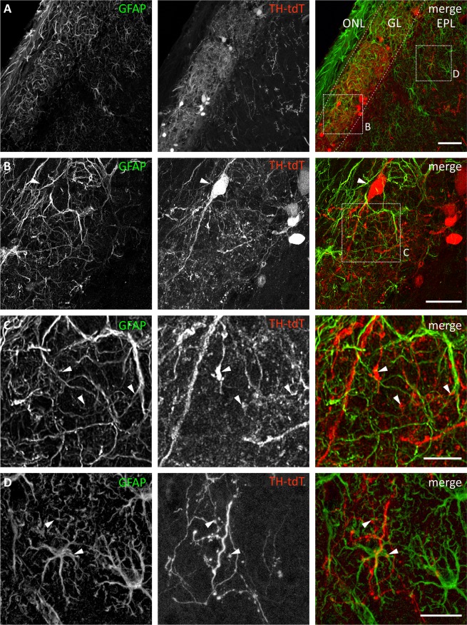
It is well established that astrocytes respond to the major neurotransmitters glutamate and GABA with cytosolic calcium rises, whereas less is known about the effect of dopamine on astroglial cells. In the present study, we used confocal calcium imaging in mouse brain slices of the olfactory bulb, a brain region with a large population of dopaminergic neurons, to investigate calcium signaling evoked by dopamine in astrocytes. Our results show that application of dopamine leads to a dose-dependent cytosolic calcium rise in astrocytes (EC50 = 76 µM) which is independent of neuronal activity and mainly mediated by PLC/IP3-dependent internal calcium release. Antagonists of both D1- and D2-class dopamine receptors partly reduce the dopaminergic calcium response, indicating that both receptor classes contribute to dopamine-induced calcium transients in olfactory bulb astrocytes.


Conflict of interest statement

The authors declare no competing interests.

Figures

Figure 1
Figure 1
Distribution of astrocytes and tyrosine hydroxylase-expressing neurons in the olfactory bulb. (A) GL, glomerular layer; ONL, olfactory nerve layer. Anti-GFAP staining (green) shows astrocytes. Cell bodies of astrocytes are located in the GL and EPL. TH+ neurons are labeled by tdTomato in THCre × tdTomatofl/fl mice (red) and are located mainly in the GL, but single neurons expressing tdTomato can be found in the EPL. Scale bar: 50 μm. (B) Magnified view from A, highlighting an interglomerular connecting short axon cell (arrowhead) in close proximity to astrocyte processes. Scale bar: 20 µm. (C) Magnified view from B, highlighting short axon cell dendrites, varicosities (arrowheads) and astrocytic processes in detail. Scale bar: 10 µm. (D) Magnified view from A, highlighting localization of astrocytes and TH+ varicosities (arrowheads). Scale bar: 20 µm.
Figure 2
Figure 2
Dopamine-triggered calcium response in olfactory bulb (OB) astrocytes. (A) Fluo-8 staining of acute OB slices with example ROIs shown in idle state (left), in the presence of 100 µM ADP (middle) and high potassium ACSF (right). (B) Example traces of cells that are considered to be a neuron (ROI1) and an astrocyte (ROI2). (C) Dopamine-induced calcium responses of astrocytes by short pressure application (500 µM, left) and bath application (100 µM, right). (D) Calcium responses of astrocytes evoked by application of ADP (30 s, 100 µM) and dopamine (DA; 30 s, 100 µM) in both EPL (black trace) and GL (blue trace). (E) Averaged amplitudes (left bars) and area (right bars) of DA-induced calcium responses in EPL and GL (error bars: SEM). Sample size as specified in the bars: cells/slices/animals. n.s.: not significant. (F) Calcium response of an astrocyte monitored during a 10-min application of DA (100 µM). (G) Calcium responses induced by application of dopamine at different concentrations. (H) Dose-response curve of dopamine-induced astroglial calcium responses (area), normalized to application of 100 µM DA (+/−SEM). (I) Percentage of astrocytes responding to dopamine application.
Figure 3
Figure 3
Dopamine-induced calcium transients in OB astrocytes in synaptic isolation. (A) Example of multiple applications of DA (100 µM) with a 10-min interval. (B) Calcium transients were not affected in presence of GABAergic, glutamatergic und purinergic antagonists (Blockermix contains: NBQX 10 µM, D-APV 50 µM, gabazine 10 µM, CGP55845 10 µM, MPEP 2 µM, MRS2179 50 µM, ZM241385 0.5 µM, TTX 1 µM). (C) Normalized averaged amplitudes (light grey, +/−SEM) and area (dark grey, +/−SEM) of calcium responses under control conditions and after application of Blockermix (BM). Results of BM are additionally compared to rundown (RD) experiment as depicted in (A) (error bars: SEM). *P < 0.05, **P < 0.01, ***P < 0.005.
Figure 4
Figure 4
Internal calcium release mediates dopamine-induced calcium transients in OB astrocytes. (A) Dopamine-induced calcium transients are entirely suppressed in the presence of SERCA inhibitor cyclopiazonic acid (CPA, 20 µM). (B) Calcium transients are partly diminished in the presence of IP3 receptor antagonist 2-APB (50 µM). (C) Attenuated calcium transients in the presence of PLC inhibitor U73122 (50 µM). (D) Normalized averaged amplitudes (left, +/−SEM) and area (right, +/−SEM) of calcium responses under control conditions and after application of CPA, 2-APB and U73122. Results are additionally compared to rundown (RD) experiment as depicted in Fig. 3A. *P < 0.05, **P < 0.01, ***P < 0.005.
Figure 5
Figure 5
Effect of DA receptor antagonists on dopamine-induced calcium signaling in OB astrocytes. (A) Calcium transients evoked by DA (30 s, 100 µM) where reduced in amplitude and integral in the presence of D1-class antagonist SCH23390 (50 µM). (B) Normalized averaged amplitude and area of calcium responses in presence of SCH23390. (C) Calcium transients where reduced in the presence of D2-class antagonist sulpiride (50 µM). (D) Normalized averaged amplitude and area of calcium responses in presence of sulpiride. (E) Effect of both SCH23390 and sulpiride on dopamine-evoked calcium responses. (F) Normalized averaged amplitude and area (+/−SEM) of calcium responses in the presence of both antagonists. *P < 0.05, **P < 0.01, ***P < 0.005.

References

    1. Schultz W. Multiple dopamine functions at different time courses. Annu. Rev. Neurosci. 2007;30:259–288. doi: 10.1146/annurev.neuro.28.061604.135722. - DOI - PubMed
    1. Felten DL, Sladek JR., Jr. Monoamine distribution in primate brain V. Monoaminergic nuclei: anatomy, pathways and local organization. Brain Res. Bull. 1983;10:171–284. doi: 10.1016/0361-9230(83)90045-x. - DOI - PubMed
    1. Cave JW, Baker H. Dopamine systems in the forebrain. Adv. Exp. Med. Biol. 2009;651:15–35. doi: 10.1007/978-1-4419-0322-8_2. - DOI - PMC - PubMed
    1. Halasz N, Johansson O, Hokfelt T, Ljungdahl A, Goldstein M. Immunohistochemical identification of two types of dopamine neuron in the rat olfactory bulb as seen by serial sectioning. J. Neurocytol. 1981;10:251–259. doi: 10.1007/BF01257970. - DOI - PubMed
    1. McLean JH, Shipley MT. Postmitotic, postmigrational expression of tyrosine hydroxylase in olfactory bulb dopaminergic neurons. J. Neurosci. 1988;8:3658–3669. doi: 10.1523/JNEUROSCI.08-10-03658.1988. - DOI - PMC - PubMed

Publication types