. 2020 Jan 20;10(1):651.
doi: 10.1038/s41598-020-57506-9.

Extracellular NAD+ enhances PARP-dependent DNA repair capacity independently of CD73 activity

Affiliations

Extracellular NAD+ enhances PARP-dependent DNA repair capacity independently of CD73 activity

Anna Wilk et al. Sci Rep. .

Abstract

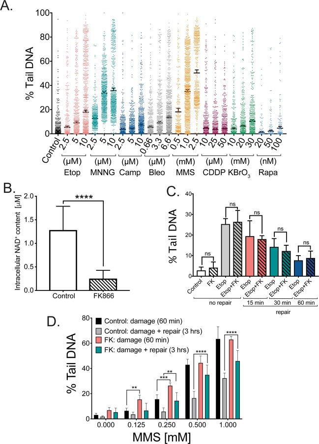
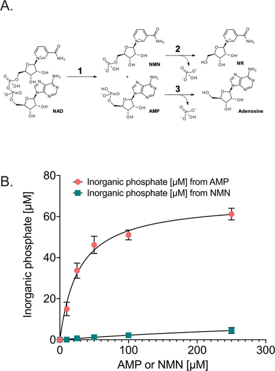
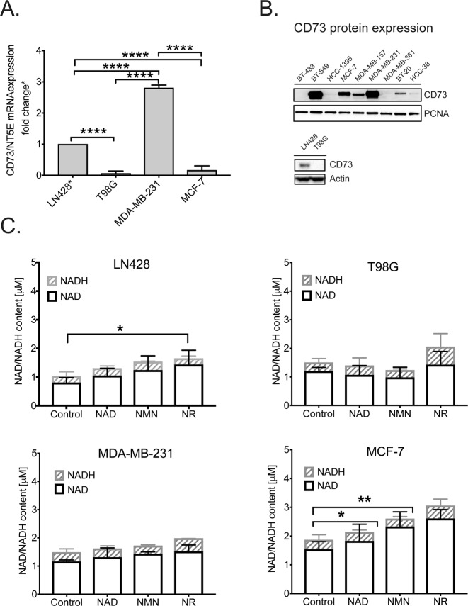
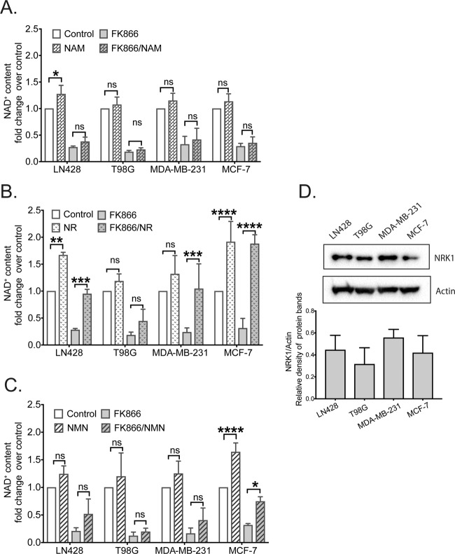
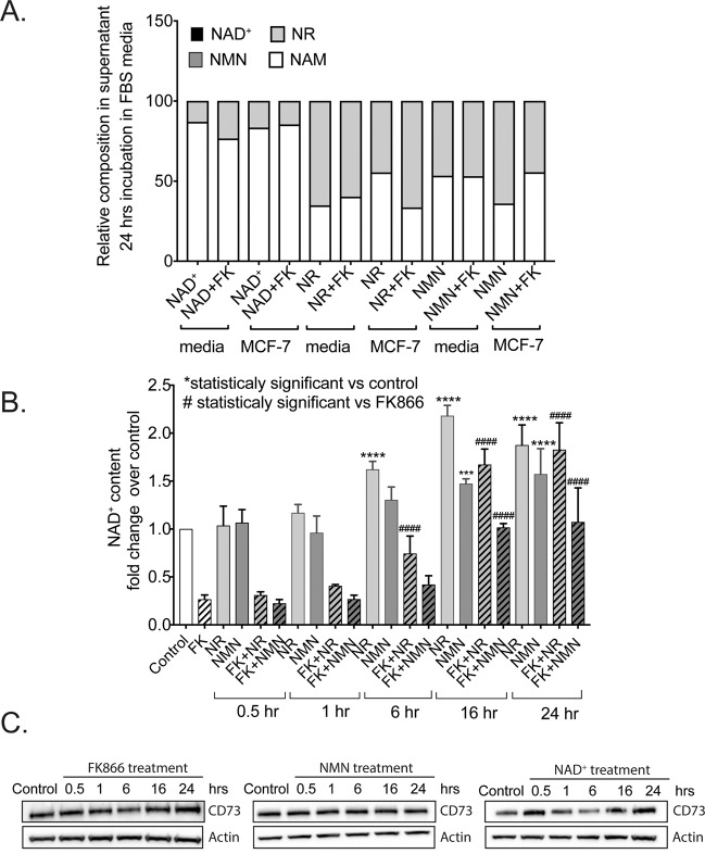
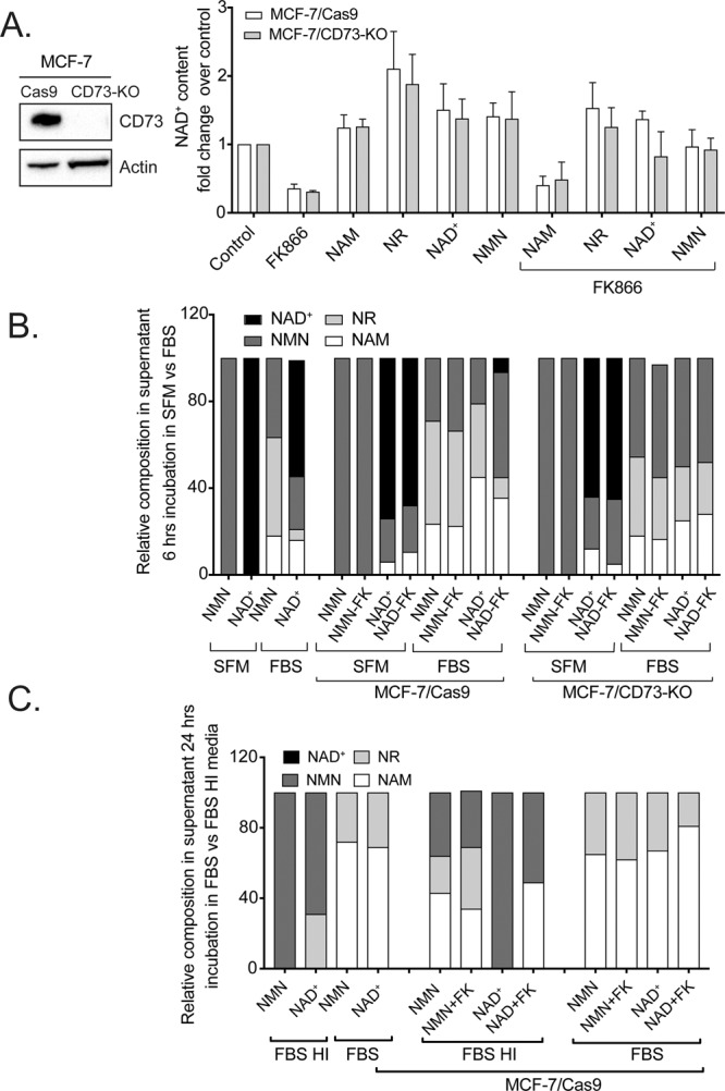
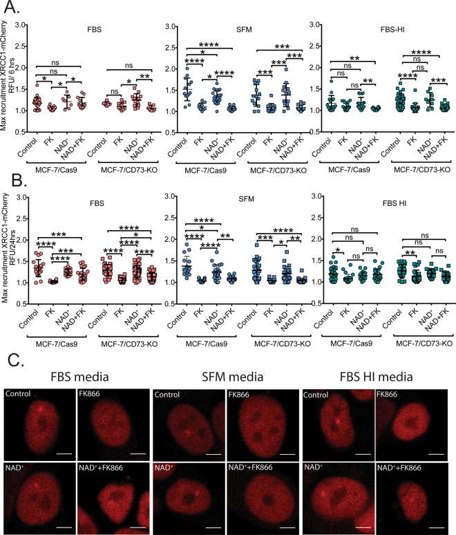
Changes in nicotinamide adenine dinucleotide (NAD+) levels that compromise mitochondrial function trigger release of DNA damaging reactive oxygen species. NAD+ levels also affect DNA repair capacity as NAD+ is a substrate for PARP-enzymes (mono/poly-ADP-ribosylation) and sirtuins (deacetylation). The ecto-5'-nucleotidase CD73, an ectoenzyme highly expressed in cancer, is suggested to regulate intracellular NAD+ levels by processing NAD+ and its bio-precursor, nicotinamide mononucleotide (NMN), from tumor microenvironments, thereby enhancing tumor DNA repair capacity and chemotherapy resistance. We therefore investigated whether expression of CD73 impacts intracellular NAD+ content and NAD+-dependent DNA repair capacity. Reduced intracellular NAD+ levels suppressed recruitment of the DNA repair protein XRCC1 to sites of genomic DNA damage and impacted the amount of accumulated DNA damage. Further, decreased NAD+ reduced the capacity to repair DNA damage induced by DNA alkylating agents. Overall, reversal of these outcomes through NAD+ or NMN supplementation was independent of CD73. In opposition to its proposed role in extracellular NAD+ bioprocessing, we found that recombinant human CD73 only poorly processes NMN but not NAD+. A positive correlation between CD73 expression and intracellular NAD+ content could not be made as CD73 knockout human cells were efficient in generating intracellular NAD+ when supplemented with NAD+ or NMN.


Conflict of interest statement

The authors declare no competing interests.

Figures

Figure 1
Figure 1
Effect of NAD+ depletion on DNA damage and repair in MCF-7 cells as determined using the CometChip assay. (A) Level of DNA damage in MCF-7 cells exposed (1 hr) to different concentrations of various DNA damaging agents. (B) Level of intracellular NAD+ in MCF-7 cells 24 hrs after treatment with FK866 (30 nM) as compared to control. Statistical analysis was conducted via an unpaired t-test (****p < 0.001). (C) Level of DNA damage and repair capacity in MCF-7 cells exposed for one hr to etoposide (10 μM) in the presence or absence of FK866 (FK; treatment with FK866 results in a 70–90% decrease in total cellular NAD+ levels). (D) Level of DNA damage and DNA repair capacity in MCF-7 cells exposed for one hr to different MMS concentrations (0, 0.125, 0.25, 0.5 and 1 mM) in the presence or absence of FK866 (FK; treatment with FK866 results in a 70–90% decrease in total cellular NAD+ levels). Data is expressed as % Tail DNA, with standard deviation. Plots are the average of 8–12 wells from two-three independent CometChips; >1000 comets per bar. Statistical analysis was performed using GraphPad Prism 7 and two-way ANOVA followed by post-hoc with Tukey’s multiple comparison test (**p < 0.0029, ***<0.0008, ****<0.0001).
Figure 2
Figure 2
Human, recombinant CD73 activity analysis for the hydrolysis of NMN, NAD+ and NADH. (A) Scheme showing the structure of NAD+ and conversion to AMP (bottom) and then to adenosine with loss of inorganic phosphate and to NMN (top) and then to NR. (B) Recombinant CD73 (rCD73; soluble form, residues 27–549 including a His-tag at the C-terminus) was purified and evaluated for enzymatic activity measured in the presence of the natural substrate, AMP, or in the presence of NMN. There was no detectable activity against NAD+ or NADH (not shown).
Figure 3
Figure 3
Comparative analysis of CD73 expression in human cancer cell lines and its effect on NAD+ biosynthesis. (A) Measurement of mRNA expression for the CD73/NT5E gene in cancer cell lines, as determined by qRT-PCR analysis, normalized to the expression of human β-actin mRNA via the ∆∆CT method. (B) Immunoblot analysis of the expression of CD73 in nine breast cancer cell lines, including the MCF-7 and MDA-MB-231 cells used herein (top panel) and two glioma cell lines, including the LN428 and T98G cells used herein (bottom panel). PCNA was used as a loading control for the top panel and actin was used as a loading control for the bottom panel. See Supplement Fig. S2F for the expression of NRK1 in the breast cancer cell lines analyzed from the same cell lysates. (C) Total intracellular NAD levels (NAD+ and NADH) in each of the four cancer cell lines cultured in the presence of NAD+, NMN or NR (100 μM) for 24 hrs. Statistical analysis was performed using GraphPad Prism 7 and two-way ANOVA followed by post-hoc test with Tukey’s correction (ns = not significant, *p = 0.0419, **p = 0.0032).
Figure 4
Figure 4
Effect of NAMPT inhibition on intracellular NAD+ content in cancer cell lines with different basal levels of CD73 protein. (A) Intracellular NAD+ levels in cells treated with FK866 (30 nM) and/or nicotinamide (NAM) (100 μM), as compared to untreated controls. (B) Intracellular NAD+ levels in cells treated with FK866 (30 nM) and/or nicotinamide riboside (NR) (100 μM), as compared to untreated controls. (C) Intracellular NAD+ levels in cells treated with FK866 (30 nM) and/or nicotinamide mononucleotide (NMN) (100 μM), as compared to untreated controls. (D) Top panel: Representative immunoblot analysis of NRK1 expression to correlate the expression of NRK1 in four cancer cell lines (see Supplement Fig. 3E for additional immunoblot figures). Bottom panel: Densitometry analysis of 3 independent immunoblots, performed using Image Lab software. Statistical analysis was performed using GraphPad Prism 7 - one or two-way ANOVA followed by post-hoc test with Tukey’s correction was used (ns = not significant, *p = 0.0176, **p = 0.0018, ***p = 0.0009 or ****p < 0.0001).
Figure 5
Figure 5
Assessment of the time dependent changes of extracellular NAD+ and NAD+ metabolite composition and intracellular NAD+ levels following metabolite supplementation and/or NAMPT inhibition. (A) NMR analysis of the composition of NAD+/NAD+-metabolites in MCF-7 cell supernatants from cells exposed to NAD+ for 24 hrs. (B) Measurements of intracellular NAD+ content in MCF-7 cells exposed to NAD supplements (NMN and NR) and/or the NAMPT inhibitor, FK866, at different time points after supplement addition. Statistical analysis was performed using GraphPad Prism 7 - one-way ANOVA followed by post-hoc test with Tukey’s correction was used. (ns =  not significant, ***p = 0.0004 or ****p < 0.0001) (C) Immunoblot analysis to evaluate changes in CD73 expression impacted by the treatment of MCF-7 cells with FK866 (30 nM), NMN (100 μM) or NAD+ (100 μM) for times ranging from 0.5 to 24 hrs, as compared to the untreated control, as indicated.
Figure 6
Figure 6
Effect of CD73 knockout on the uptake of NAD+ precursors and on NAD+ biosynthesis. (A) Left panel: Immunoblot analysis of CD73 expression in MCF-7/Cas9 and MCF-7/CD73-KO cells; actin is shown as a loading control. Right panel: Comparative analysis of intracellular NAD+ levels in both cell lines exposed to different NAD+ precursors (100 μM) in the absence or presence of the NAMPT inhibitor, FK866 (30 nM). (B) NMR analysis to define the distribution of NAD+ and NAD+ metabolites (100 μM) in cell culture media 6 hrs after supplementation of NMN or NAD+ to cell-free serum-free media (SFM), cell-free media supplemented with fetal bovine serum (FBS) or when added to SFM or FBS in the presence of MCF-7/Cas9 or MCF-7/CD73-KO cells in the presence or absence of FK866 (30 nM). (C) NMR analysis to define the distribution of NAD+ and NAD+ metabolites in cell culture media 24 hrs after supplementation of NMN or NAD+ to MCF-7/Cas9 cells in the presence or absence of FK866. Cells were exposed to media supplemented with fetal bovine serum (FBS) or heat inactivated fetal bovine serum (FBS-HI) and compared to NMN and NAD+, which have never been exposed to cells.
Figure 7
Figure 7
Impact of NAD+ depletion and NAD+ precursor supplementation on DNA repair complex formation. (A, B) Comparative measurements of maximal recruitment of XRCC1-mCherry protein to the site of laser-induced DNA damage: MCF-7/Cas9 vs MCF-7/CD73-KO cells exposed to NAD+ in the presence of serum-free media (SFM) or media supplemented with fetal bovine serum (FBS) or heat inactivated fetal bovine serum (FBS-HI) for 6 hrs (A) or 24 hrs (B). (C) Representative images of maximal recruitment of XRCC1-mCherry in MCF-7/Cas9 cells cultured in media+FBS, SFM or media+FBS-HI; respectively, with differing NAD+ status.

References

    1. Kepp O, Loos F, Liu P, Kroemer G. Extracellular nucleosides and nucleotides as immunomodulators. Immunol. Rev. 2017;280:83–92. doi: 10.1111/imr.12571. - DOI - PubMed
    1. Wilde L, et al. Metabolic coupling and the R everse Warburg Effect in cancer: Implications for novel biomarker and anticancer agent development. Semin. Oncol. 2017;44:198–203. doi: 10.1053/j.seminoncol.2017.10.004. - DOI - PMC - PubMed
    1. Otto AM, Hintermair J, Janzon C. NADH-linked metabolic plasticity of MCF-7 breast cancer cells surviving in a nutrient-deprived microenvironment. J. Cell Biochem. 2015;116:822–835. doi: 10.1002/jcb.25038. - DOI - PubMed
    1. Thongon N, et al. Cancer cell metabolic plasticity allows resistance to NAMPT inhibition but invariably induces dependence on LDHA. Cancer Metab. 2018;6:1. doi: 10.1186/s40170-018-0174-7. - DOI - PMC - PubMed
    1. Bogan KL, Brenner C. Nicotinic acid, nicotinamide, and nicotinamide riboside: a molecular evaluation of NAD+ precursor vitamins in human nutrition. Annu. Rev. Nutr. 2008;28:115–130. doi: 10.1146/annurev.nutr.28.061807.155443. - DOI - PubMed

Publication types

MeSH terms