. 2020 Feb;21(2):178-185.
doi: 10.1038/s41590-019-0578-8. Epub 2020 Jan 20.

Genome-wide CRISPR-Cas9 screening reveals ubiquitous T cell cancer targeting via the monomorphic MHC class I-related protein MR1

Affiliations

Genome-wide CRISPR-Cas9 screening reveals ubiquitous T cell cancer targeting via the monomorphic MHC class I-related protein MR1

Michael D Crowther et al. Nat Immunol. 2020 Feb.

Erratum in

Abstract

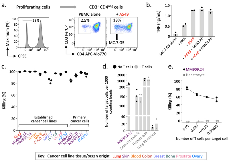
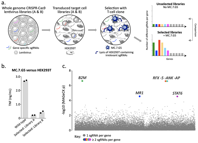
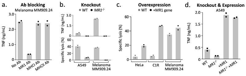
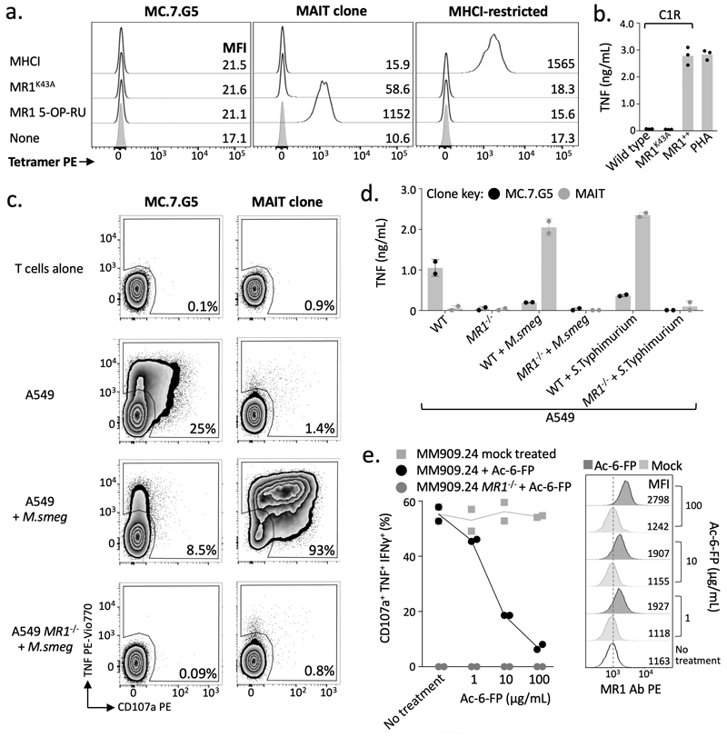
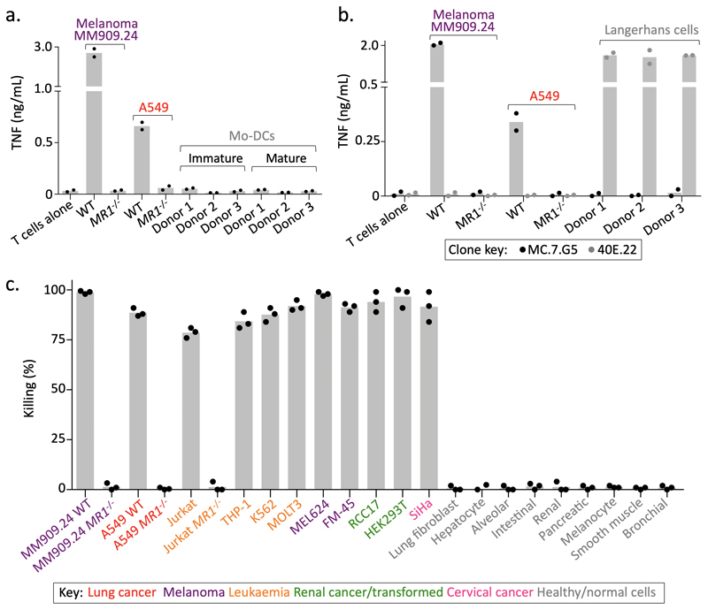
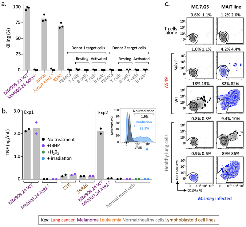
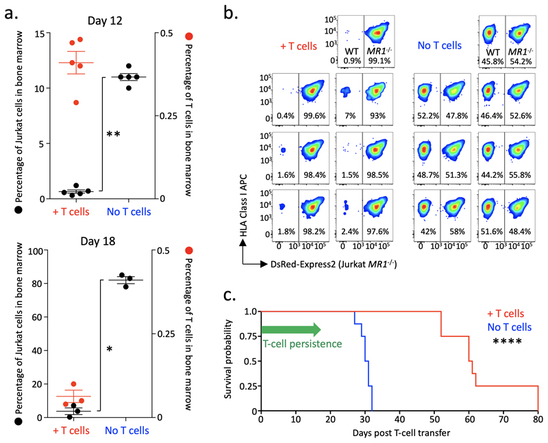
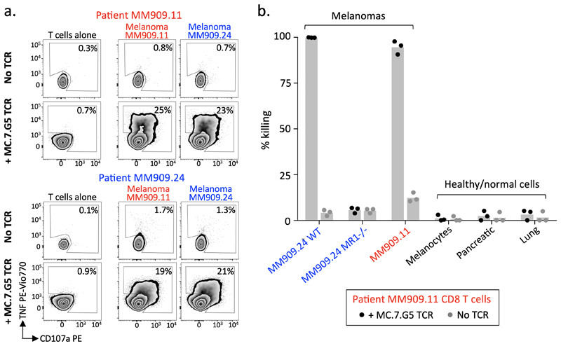
Human leukocyte antigen (HLA)-independent, T cell-mediated targeting of cancer cells would allow immune destruction of malignancies in all individuals. Here, we use genome-wide CRISPR-Cas9 screening to establish that a T cell receptor (TCR) recognized and killed most human cancer types via the monomorphic MHC class I-related protein, MR1, while remaining inert to noncancerous cells. Unlike mucosal-associated invariant T cells, recognition of target cells by the TCR was independent of bacterial loading. Furthermore, concentration-dependent addition of vitamin B-related metabolite ligands of MR1 reduced TCR recognition of cancer cells, suggesting that recognition occurred via sensing of the cancer metabolome. An MR1-restricted T cell clone mediated in vivo regression of leukemia and conferred enhanced survival of NSG mice. TCR transfer to T cells of patients enabled killing of autologous and nonautologous melanoma. These findings offer opportunities for HLA-independent, pan-cancer, pan-population immunotherapies.


Conflict of interest statement

Competing Interests

Cardiff University has filed patents based on these findings.

Figures

Figure 1
Figure 1. MC.7.G5 recognises multiple cancer types through an HLA-independent mechanism.
(a) MC.7.G5 was cloned from T cells that proliferated in response to cancer cell line A549. Performed once for this donor. (b) MC.7.G5 did not recognise A549 through MHCI or MHCII. Overnight activation +/- blocking antibodies and TNF ELISA. Bars depict the mean. (c) MC.7.G5 killed a range of established (long-term culture) and primary cancer cell lines of different origin. Flow-based killing assay for 48-72h at a T cell to target cell ratio of 5:1. Data combined from different experiments. Performed in triplicate. (d) MC.7.G5 killed melanoma cells but not healthy cells. Flow based killing assay at a T cell to target cell ratio of 5:1. Performed in triplicate or duplicate (fibroblasts). (e) MC.7.G5 sensitively killed melanoma MM909.24 over 7 days. Performed in duplicate. b-e Bars, horizontal lines and connecting line depict the mean.
Figure 2
Figure 2. Whole genome CRISPR-Cas9 library screening reveals MR1 as the candidate target of MC.7.G5.
(a) Overview of the approach used to reveal the ligand of MC.7.G5. GeCKO v2 whole genome CRISPR-Cas9 libraries A and B were used as lentivirus to transduce target cell line HEK293T. MC.7.G5 lysed HEK293T expressing sgRNAs for genes that are irrelevant for HEK293T recognition, thereby enriching sgRNAs for genes that are essential for cancer cell lysis by MC.7.G5. Two rounds of selection with MC.7.G5 were performed and comparison of selected libraries unselected HEK293T (no MC.7.G5) revealed enriched sgRNAs. (b) MC.7.G5 recognition of selected HEK293T library post-selection is greatly reduced compared to wild-type HEK293T, suggesting key genes had been ablated by the whole genome CRISPR-Cas9 approach. Overnight activation and TNF ELISA, performed in duplicate. Bars depict the mean. (c) MR1 was identified as one of key genes for MC.7.G5 recognition of HEK293T. Total genomic DNA from 3 x10⁷ selected and unselected HEK293T libraries was used for sequencing, followed by MAGeCK analysis. Highlighted (coloured) genes with y genes for MC.7.G5 recognition of HEK293T.reduced
Figure 3
Figure 3. MR1 is the cancer cell expressed target of MC.7.G5.
(a) Recognition of melanoma MM909.24 was reduced in the presence of MR1 blocking antibody (Ab). MHCI and II Abs were used as negative controls. Overnight activation and TNF ELISA. (b) Removal of MR1 expression (CRISPR/Cas9) from cancer cell lines prevented MC.7.G5 mediated recognition and killing. Overnight activation and TNF ELISA or chromium release cytotoxicity assay. (c) Lentiviral overexpression (+) of MR1 in poor targets of MC.7.G5 improved target cell killing by MC.7.G5. Chromium release cytotoxicity assay. (d) Lentiviral expression of MR1 in MR1-/- cells restores activation of MC.7.G5. TNF ELISA. Conditions performed in duplicate. Bars depict the mean.
Figure 4
Figure 4. MC.7.G5 does not recognise MR1 by known mechanisms.
(a) MC.7.G5 did not stain with empty (K43A) or MR1 5-OP-RU tetramers. A canonical MAIT clone recognizes MR1 bound with 5-OP-RU. The MHCI-restricted clone was used as a positive control for the irrelevant MHCI tetramer. Performed twice with similar results. (b) MC.7.G5 recognised target cells over-expressing wildtype MR1 (MR1++) but not K43A mutated MR1. Overnight activation performed in duplicate and TNF ELISA. (c) Loading with MAIT-activating bacterium Mycobacterium smegmatis (M.smeg) reduced MC.7.G5 recognition of A549 cells. Canonical MAIT clone used as a positive control. Staining for surface CD107a and intracellular TNF. Performed twice with similar results. (d) M. smeg and Salmonella enterica serovar Typhimurium (S.Typhimurium) reduced MC.7.G5 recognition of A549 cells. Overnight activation and TNF ELISA. (e) Exogenous Ac-6-FP, a known MR1 binding molecule, reduced MC.7.G5 recognition of melanoma MM909.24. Percentage of cell triple positive for the markers shown is plotted. Performed twice with similar results.
Figure 5
Figure 5. MC.7.G5 does not recognise healthy cells.
(a) MC.7.G5 did not recognize immature or matured monocyte (mo) derived dendritic cells (DCs). Overnight activation and TNF ELISA. (b) MC.7.G5 did not recognize matured Langerhans cells. CD1a-restricted clone 40E.22 used as a positive control for recognition of Langerhans cells. Overnight activation and TNF ELISA. (c) Cancer cell lines lacking MR1 (CRISPR/Cas9) and healthy cells from various tissues were not killed by MC.7.G5. Flow-based killing assay (48h 1:1 ratio). Performed in triplicate. a-c bars depict the mean.
Figure 6
Figure 6. MC.7.G5 remained inert to activated, stressed or infected healthy cells.
(a) T cell (Jurkat) and B cell (K562) cancer cells were targets of MC.7.G5, whereas whole PBMCs and resting or activated purified T and B cells were not killed. Flow-based killing assay (24h 1:1 ratio). Performed in triplicate. (b) Experiment 1: tert-Butyl hydroperoxide (tBHP) treatment to induce stress in poor targets (C1R and SAR26 lymphoblastoid cell lines) of MC.7.G5 did not lead to T cell activation. MC.7.G5 recognition of melanoma MM909.24 +/- MR1 was unaffected by tBHP treatment. Experiment 2: Healthy renal epithelial cells were not recognised by MC.7.G5 following treatment with either tBHP or hydrogen peroxide (H202), or after exposure to γ-irradiation. Overnight activation and TNF ELISA. Inserted histogram of irradiated renal cells stained with the viability dye VIVID showing cell death after irradiation compared to un-irradiated cells. (c) Mycobacterium smegmatis infected healthy lung epithelial cells did not lead to MC.7.G5 activation, whereas a MAIT line recognised the infected cells. Uninfected or infected A459 cells +/- MR1 acted as controls for MC.7.G5 and the MAIT line respectively. The MAIT line exhibited some recognition towards the uninfected lung cells. TAPI-0 assay for 4h. Percentage shown for duplicate conditions. Performed twice with similar results. a-c bars depict the mean.
Figure 7
Figure 7. MC.7.G5 mediates in vivo regression of leukemia and prolongs the survival of mice.
(a) NSG mice received Jurkat cells (3 x10⁶) then a single infusion of MC.7.G5 (1.5 x10⁶) 7 days later. MC.7.G5 reduced Jurkat cells in bone marrow cells at day 12 (n=10) and 18 (n=6) post T cell transfer (left axis). P values (* 0.032 ** 0.0038) from a two-sided non-parametric two-sample Kolmogorov-Smirnov test. Horizontal line depicts mean and error bars the SD. Jurkat cells did not appear in the spleen at day 12 but MC.7.G5 reduced Jurkat cell load by day 18. Few MC.7.G5 cells were recovered from the bone marrow 18 days after single infusion (right y-axis), and also from the spleen. (b) WT MR1 expressing Jurkat cells were preferentially targeted in mice receiving MC.7.G5. The same number of MR1 WT and MR1-/- (DsRed-Express2+) Jurkat cells (4 x10⁶ in total) were co-transferred to the same mouse (n=7 per group) followed 7 days later by 3 x10⁶ MC.7.G5. Splenocytes were harvested on day d25 post T cell transfer. (c) Enhanced survival of mice with Jurkat cancer that received MC.7.G5. 8 mice per group (+/- T cells). Experimental set-up as in (a). Mice were culled when they had lost 15% of their original body weight as required by UK Home Office rules. Median survival of 60.5 and 30.5 days for +/- T cells respectively. Logrank two-sided p value (**** 0.000066) and Hazard Ratio (4.54, 1.27-16.21) were calculated using the MatSurv survival analysis function in Matlab, availableilabhttps://www.github.com/aebergl/MatSurv.
Figure 8
Figure 8. Transfer of the MC.7.G5 T cell receptor redirects patient T cells to recognise autologous melanoma.
(a) Metastatic melanoma patient (MM909.11 and MM909.24) derived T cells transduced with the T cell receptor of MC.7.G5 recognised autologous and non-autologous melanomas. Surface CD107a and intracellular TNF after 4h. Performed twice with similar results. (b) T cells from patient MM909.11 transduced with MC.7.G5 TCR killed autologous and non-autologous melanomas, but not healthy cells. Flow-based killing assay for 36h at a T cell to target cell ratio of 5:1. Bars depict the mean.

Comment in

  • 'Bohemian Rhapsody' of MR1T cells.
    Mori L, De Libero G. Mori L, et al. Nat Immunol. 2020 Feb;21(2):108-110. doi: 10.1038/s41590-019-0588-6. Nat Immunol. 2020. PMID: 31959981 No abstract available.
  • MR1-restricted pan-cancer T cells.
    Bird L. Bird L. Nat Rev Immunol. 2020 Mar;20(3):141. doi: 10.1038/s41577-020-0284-7. Nat Rev Immunol. 2020. PMID: 32024985 No abstract available.

References

    1. Vavassori S, et al. Butyrophilin 3A1 binds phosphorylated antigens and stimulates human γδ T cells. Nat Immunol. 2013;14:908–916. - PubMed
    1. Kjer-Nielsen L, et al. MR1 presents microbial vitamin B metabolites to MAIT cells. Nature. 2012;491:717–723. - PubMed
    1. Corbett AJ, et al. T-cell activation by transitory neo-antigens derived from distinct microbial pathways. Nature. 2014;509:361–5. - PubMed
    1. Gold MC, et al. MR1-restricted MAIT cells display ligand discrimination and pathogen selectivity through distinct T cell receptor usage. J Exp Med. 2014;211:1601–1610. - PMC - PubMed
    1. Eckle SBG, et al. Recognition of Vitamin B precursors and byproducts by mucosal associated invariant T cells. J Biol Chem. 2015;290:30204–30211. - PMC - PubMed

Publication types

MeSH terms

Substances