

PEBP1/RKIP behavior: a mirror of actin-membrane organization
- PMID: 31960115
- PMCID: PMC11105014
- DOI: 10.1007/s00018-020-03455-5
PEBP1/RKIP behavior: a mirror of actin-membrane organization
Abstract
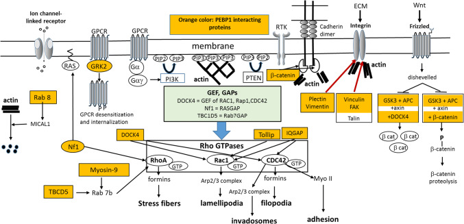
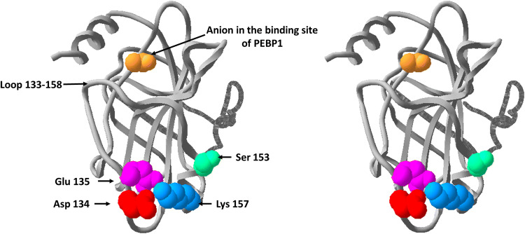
Phosphatidylethanolamine-binding protein 1 (PEBP1), a small 21 kDa protein, is implicated in several key processes of the living cell. The deregulation of PEBP1, especially its downregulation, leads to major diseases such as cancer and Alzheimer's disease. PEBP1 was found to interact with numerous proteins, especially kinases and GTPases, generally inhibiting their activity. To understand the basic functionality of this amazing small protein, we have considered several known processes that it modulates and we have discussed the role of each molecular target in these processes. Here, we propose that cortical actin organization, associated with membrane changes, is involved in the majority of the processes modulated by PEBP1. Furthermore, based on recent data, we summarize some key PEBP1-interacting proteins, and we report their respective functions and focus on their relationships with actin organization. We suggest that, depending on the cell status and environment, PEBP1 is an organizer of the actin-membrane composite material.
Keywords: Actin; Cell shape; Cytoskeleton; Membrane; Motility.
Figures
References
-
- Bernier I, Tresca JP, Jollès P. Ligand-binding studies with a 23 kDa protein purified from bovine brain cytosol. Biochim Biophys Acta. 1986;871(1):19–23. - PubMed
-
- Schoentgen F, Saccoccio F, Jollès J, Bernier I, Jollès P. Complete amino acid sequence of a basic 21-kDa protein from bovine brain cytosol. Eur J Biochem. 1987;166(2):333–338. - PubMed
-
- Serre L, Vallée B, Bureaud N, Schoentgen F, Zelwer C. Crystal structure of the Phosphatidylethanolamine-binding protein from bovine brain: a novel structural class of phospholipid-binding proteins. Structure. 1998;6(10):1255–1265. - PubMed
-
- Banfield MJ, Barker JJ, Perry AC, Brady RL. Function from structure? The crystal structure of human phosphatidylethanolamine-binding protein suggests a role in membrane signal transduction. Structure. 1998;6(10):1245–1254. - PubMed
-
- Lamiman K, Keller JM, Mizokami A, Zhang J, Keller ET. Survey of Raf kinase inhibitor protein (RKIP) in multiple cancer types. Crit Rev Oncog. 2014;19(6):455–468. - PubMed
MeSH terms
Substances
LinkOut - more resources
Full Text Sources
Research Materials
Miscellaneous
