. 2020 Mar;37(3):525-542.
doi: 10.1007/s12640-020-00164-6. Epub 2020 Jan 21.

Neuroprotective Effects of Necrostatin-1 Against Oxidative Stress-Induced Cell Damage: an Involvement of Cathepsin D Inhibition

Affiliations

Neuroprotective Effects of Necrostatin-1 Against Oxidative Stress-Induced Cell Damage: an Involvement of Cathepsin D Inhibition

Danuta Jantas et al. Neurotox Res. 2020 Mar.

Abstract

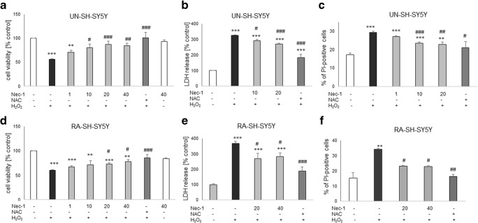
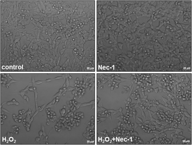
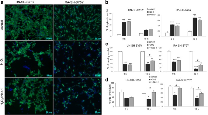
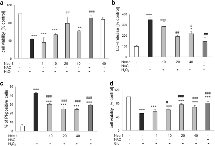
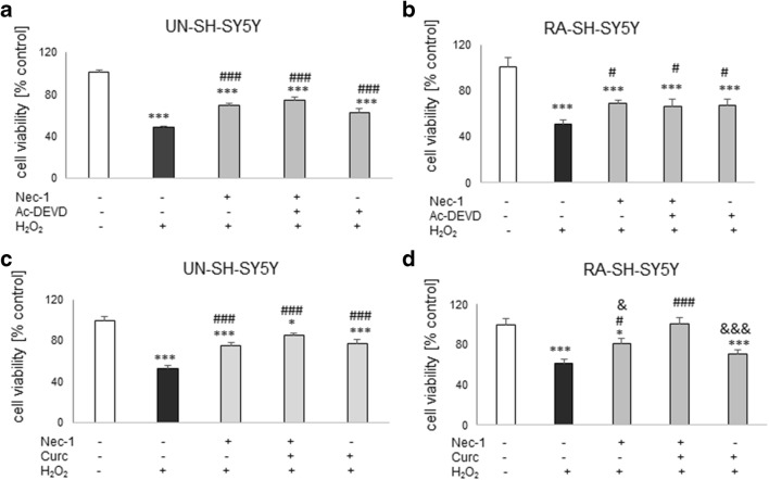
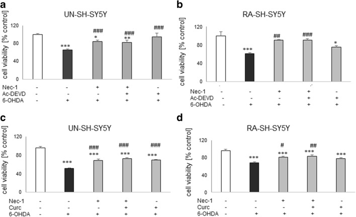
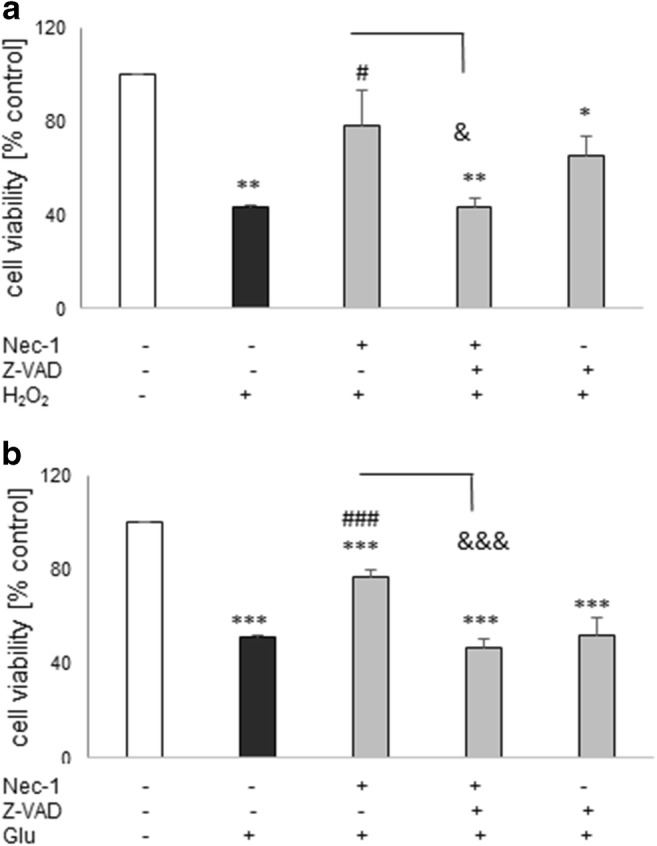
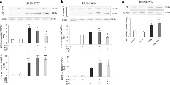
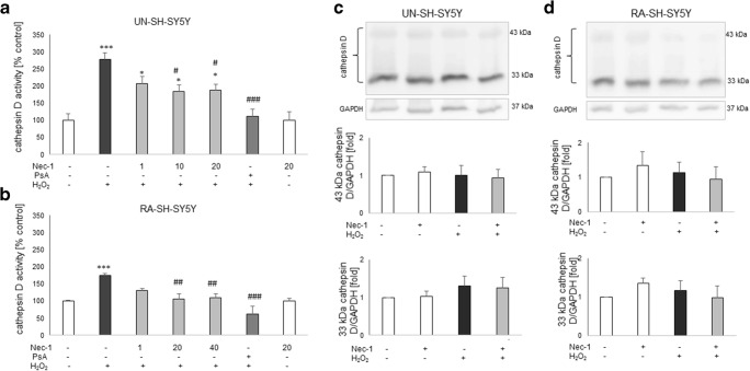
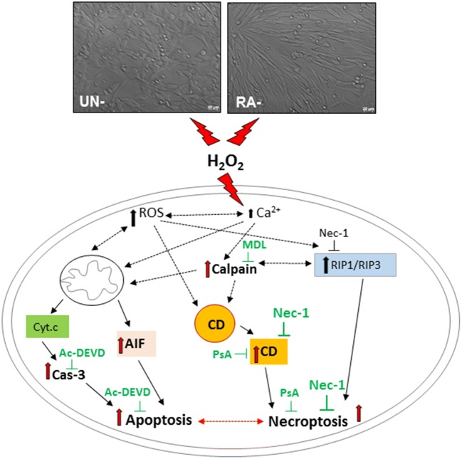
Necroptosis, a recently discovered form of non-apoptotic programmed cell death, can be implicated in many pathological conditions including neuronal cell death. Moreover, an inhibition of this process by necrostatin-1 (Nec-1) has been shown to be neuroprotective in in vitro and in vivo models of cerebral ischemia. However, the involvement of this type of cell death in oxidative stress-induced neuronal cell damage is less recognized. Therefore, we tested the effects of Nec-1, an inhibitor of necroptosis, in the model of hydrogen peroxide (H2O2)-induced cell damage in human neuroblastoma SH-SY5Y and murine hippocampal HT-22 cell lines. The data showed that Nec-1 (10-40 μM) attenuated the cell death induced by H2O2 in undifferentiated (UN-) and neuronal differentiated (RA-) SH-SY5Y cells with a higher efficacy in the former cell type. Moreover, Nec-1 partially reduced cell damage induced by 6-hydroxydopamine in UN- and RA-SH-SY5Y cells. The protective effect of Nec-1 was of similar magnitude as the effect of a caspase-3 inhibitor in both cell phenotypes and this effect were not potentiated after combined treatment. Furthermore, the non-specific apoptosis and necroptosis inhibitor curcumin augmented the beneficial effect of Nec-1 against H2O2-evoked cell damage albeit only in RA-SH-SY5Y cells. Next, it was found that the mechanisms of neuroprotective effect of Nec-1 against H2O2-induced cell damage in SH-SY5Y cells involved the inhibition of lysosomal protease, cathepsin D, but not caspase-3 or calpain activities. In HT-22 cells, Nec-1 was protective in two models of oxidative stress (H2O2 and glutamate) and that effect was blocked by a caspase inhibitor. Our data showed neuroprotective effects of the necroptosis inhibitor, Nec-1, against oxidative stress-induced cell damage and pointed to involvement of cathepsin D inhibition in the mechanism of its action. Moreover, a cell type-specific interplay between necroptosis and apoptosis has been demonstrated.

Keywords: Caspase-3; Glutamate; HT-22 cells; Hydrogen peroxide; Pepstatin A; SH-SY5Y cells.


Conflict of interest statement

The authors declare that they have no conflicts of interest.

Figures

Fig. 1
Fig. 1
The effect of necrostatin-1 on H2O2-induced cell damage in UN- and RA-SH-SY5Y cells. UN- and RA-SH-SY5Y cells (ac and df, respectively) were pre-treated for 30 min with necrostatin-1 (Nec-1; 1–40 μM) followed by 24 h of treatment with H2O2 (0.25 and 0.5 mM for UN- and RA-SH-SY5Y, respectively). As a positive control for the assays, we used antioxidant N-acetylcysteine (NAC, 1 mM) which was given concomitantly with the cell damaging factor. a, d Results of cell viability assessment in UN-(a) and RA-(d) SH-SY5Y cells measured by the MTT reduction assay. Data were normalized to vehicle-treated cells (control) and are presented as the mean ± SEM from 3 to 11 separate experiments with 5 repetitions each. (b, e) Results of cell toxicity assessment in UN-(b) and RA-(e) SH-SY5Y cells measured by the LDH release assay. Data were normalized to vehicle-treated cells (control) and are presented as the mean ± SEM from 4 to 11 separate experiments with 5 repetitions each. c, f Flow cytometry results of propidium iodide (PI)-stained UN- (c) and RA (f) SH-SY5Y cells after 24 h of cell treatment. Data are presented as the mean ± SEM of PI-positive cells from 3 to 5 independent experiments with 2 replicates. **P < 0.01 and ***P < 0.001 vs. vehicle-treated cells; #P < 0.05, ##P < 0.01, and ###P < 0.001 vs. H2O2-treated cells
Fig. 2
Fig. 2
Representative DIC (differential interference contrast) images of UN-SH-SY5Y cells treated for 24 h with necrostatin-1 (Nec-1, 20 μM) and hydrogen peroxide (H2O2, 0.25 mM)
Fig. 3
Fig. 3
a Representative microphotographs of UN- and RA-SH-SY5Y cells double-stained with CalceinAM/Hoechst 33342 after 18 h of treatment with necrostatin-1 (Nec-1; 20 μM) and hydrogen peroxide (H2O2; 0.25 and 0.5 mM for UN- and RA-SH-SY5Y cells, respectively). b, c An estimation of number of pyknotic (b) and healthy nuclei (c) from Hoechst 33342 staining. Nuclei showing bright blue florescence (condensed or fragmented) staining were semi-manually counted and presented as the mean percentage of pyknotic nuclei/all nuclei ± SEM or percentage of healthy nuclei (normalized to control group) from two independent experiments with two replicates. d Quantification of neurite length (in μm) from CalceinAM staining using Simple Neurite Tracer (an ImageJ add-on software). The lengths of ten random neurites per image, 3 images per well, and 2 wells per experimental group were measured. The data are presented as a mean neurite length ± SEM (in μm) from 2 independent experiments. Statistical analysis was performed with one-way ANOVA with Tukey’s post hoc test independently for each time point. ***P < 0.05, **P < 0.01, and ***P < 0.001 vs. vehicle-treated cells; #P < 0.05 vs. H2O2-treated cells
Fig. 4
Fig. 4
The effect of necrostatin-1 on H2O2- (ac) or glutamate- (Glu, d) induced cell damage in hippocampal HT-22 cells. The cells were pre-treated for 30 min with necrostatin-1 (Nec-1; 1–40 μM) followed by 24 h of treatment with H2O2 (1 mM) or Glu (3 mM). As a positive control for the assays, we used antioxidant N-acetylcysteine (NAC, 1 mM) which was given concomitantly with the cell damaging factors. a, d Results of cell viability assessment in the model of cell damage induced by H2O2- (a) and Glu (d) measured by the MTT reduction assay. Data were normalized to vehicle-treated cells (control) and are presented as the mean ± SEM from 3 to 10 separate experiments with 5 repetitions each. b Results of cell toxicity assessment in HT-22 cell exposed to H2O2 and Nec-1 measured by the LDH release assay. Data were normalized to vehicle-treated cells (control) and are presented as the mean ± SEM from 3 to 5 separate experiments with 5 repetitions each. c Flow cytometry results of propidium iodide (PI)-stained HT-22 cells after 24 h of cell treatment with H2O2 and Nec-1. Data are presented as the mean ± SEM of PI-positive cells from 3 to 4 independent experiments with 2 replicates. *P < 0.05, **P < 0.01, and ***P < 0.001 vs. vehicle-treated cells; #P < 0.05, ##P < 0.01, and ###P < 0.001 vs. H2O2- or Glu-treated cells
Fig. 5
Fig. 5
The effect of combined treatment with necrostatin-1 (Nec-1) and caspase-3 inhibitor (Ac-DEVD-CHO) (a, b) or Nec-1 and curcumin (Curc) against the hydrogen peroxide-induced cell damage in UN- (a, c) and RA- (b, d) SH-SY5Y cells. The cells were pre-treated for 30 min with Nec-1 (20 μM) and Ac-DEVD-CHO (20 μM) or Nec-1 (20 μM) and Curc (5 μM) alone or in combination followed by 24 h of treatment with H2O2 (0.25 mM and 0.5 mM for UN- and RA-SH-SY5Y cells). Cell viability was estimated by the MTT reduction assay, and data were normalized to vehicle-treated cells (control) and are presented as the mean ± SEM from 3 separate experiments with 5 repetitions each. *P < 0.05 and ***P < 0.001 vs. vehicle-treated cells; #P < 0.05 and ###P < 0.001 vs. H2O2-treated cells; &P < 0.05 and &&&P < 0.001 vs. H2O2 + Nec-1 + Curc-treated cells
Fig. 6
Fig. 6
The effect of combined treatment with necrostatin-1 (Nec-1) and caspase-3 inhibitor (Ac-DEVD-CHO) (a, b) or Nec-1 and curcumin (Curc) (c, d) against the 6-OHDA-induced cell damage in UN- (a, c) and RA- (b, d) SH-SY5Y cells. The cells were pre-treated for 30 min with Nec-1 (20 μM) and Ac-DEVD-CHO (20 μM) or Nec-1 (20 μM) and Curc (5 μM) alone or in combination followed by 24 h of treatment with 6-OHDA (0.1 and 0.2 mM for UN- and RA-SH-SY5Y cells, respectively). Cell viability was estimated by the MTT reduction assay, and data were normalized to vehicle-treated cells (control) and are presented as the mean ± SEM from 3 separate experiments with 5 repetitions each. *P < 0.05, **P < 0.01, and ***P < 0.001 vs. vehicle-treated cells; #P < 0.05, ##P < 0.01, and ###P < 0.001 vs. H2O2-treated cells
Fig. 7
Fig. 7
The effect of combined treatment with necrostatin-1 (Nec-1) and pan-caspase inhibitor (Z-VAD-fmk) against the H2O2- (a) and glutamate- (Glu, b) induced cell damage in HT-22 cells. The cells were pre-treated for 30 min with Nec-1 (20 μM) and Z-VAD-fmk (20 μM) alone or in combination followed by 24 h of treatment with H2O2 (1 mM) or Glu (3 mM). Cell viability was estimated by the MTT reduction assay, and data were normalized to vehicle-treated cells (control) and are presented as the mean ± SEM from 3 to 7 separate experiments with 5 repetitions each. *P < 0.05, **P < 0.01, and ***P < 0.001 vs. vehicle-treated cells; #P < 0.05 and ###P < 0.001 vs. H2O2- or Glu-treated cells; &P < 0.05 and &&&P < 0.001 vs. H2O2 + Nec-1 or Glu + Nec-1-treated cells
Fig. 8
Fig. 8
a, b The effect of necrostatin-1 (Nec-1) on the H2O2-induced 145 kDa and 120 kDa spectrin α II breakdown products in UN- and RA-SH-SY5Y cells, which are specifically cleaved by calpains and caspases, respectively. Cells were pre-treated for 30 min with Nec-1 (20 μM) or calpain inhibitor MDL28170 (10 μM), followed by 14 h of treatment with H2O2 (0.25 and 0.5 mM for UN- and RA-SH-SY5Y cells, respectively). c The effect of Nec-1 on the H2O2-induced increase in cytosolic AIF (apoptosis inducing factor) level. The UN-SH-SY5Y cells were pre-treated for 30 min with Nec-1 (20 μM) followed by 14 h of treatment with H2O2 (0.25 mM). ac Histograms show the quantified Western blot results from duplicate determinations in 2–3 independent experiments which were normalized to the protein loading control (GAPDH) and are expressed as fold of the control ± SEM. *P < 0.05, **P < 0.01, and ***P < 0.001 vs. vehicle-treated cells; #P < 0.05 vs. H2O2-treated cells
Fig. 9
Fig. 9
a, b The effect of necrostatin-1 (Nec-1) on H2O2-induced cathepsin D activity in UN- (a) and RA (b) SH-SY5Y cells. The cells were pre-treated for 30 min with Nec-1 (1–40 μM) or pepstatin A (PsA; 0.2 μM) followed by 18 h of treatment with H2O2 (0.25 and 0.5 mM for UN- and RA-SH-SY5Y cells, respectively). Data from duplicate determinations in 3–4 independent experiments were normalized to the protein level and are expressed as percentages of the control ± SEM. c, d The effect of necrostatin-1 (Nec-1) on H2O2-induced cathepsin D expression in UN- (c) and RA- (d) SH-SY5Y cells. The cells were pre-treated for 30 min with Nec-1 (20 μM) followed by 18 h of treatment with H2O2 (0.25 and 0.5 mM for UN- and RA-SH-SY5Y cells, respectively). Expression of 43 and 33 kDa forms of cathepsin D was done by Western blot method. Histograms show the quantified WB results from duplicate determinations in 2 independent experiments which were normalized to the protein loading control (GAPDH) and are expressed as fold of the control ± SEM
Fig. 10
Fig. 10
A schematic illustration of possible mechanisms by which the necrostatin-1 (Nec-1) mediates neuroprotection against the H2O2-induced cell damage in undifferentiated (UN-) and retinoic acid (RA)-differentiated SHSY5Y cells. Cathepsin D inhibition, but not caspase-3, calpain or AIF (apoptosis inducing factor) translocation inhibition, is proposed as a candidate mechanism associated with neuroprotection mediated by Nec-1 in oxidative stress (H2O2)-evoked cell damage. Ac-DEVD—Ac-DEVD-CHO, an inhibitor of caspase-3; MDL—MDL28170, an inhibitor of calpains; CD—cathepsin D; PsA—pepstatin A, an inhibitor of cathepsin D; H2O2—hydrogen peroxide; ROS—reactive oxygen species

Similar articles

Cited by

References

    1. Agholme L, Lindström T, Kågedal K, Marcusson J, Hallbeck M. An in vitro model for neuroscience: differentiation of SH-SY5Y cells into cells with morphological and biochemical characteristics of mature neurons. J Alzheimers Dis. 2010;20:1069–1082. - PubMed
    1. Arrázola MS, Saquel C, Catalán RJ, Barrientos SA, Hernandez DE, Martínez NW, Catenaccio A, Court FA. Axonal degeneration is mediated by necroptosis activation. J Neurosci. 2019;39:3832–3844. - PMC - PubMed
    1. Askalan R, Gabarin N, Armstrong EA, Fang Liu Y, Couchman D, Yager JY. Mechanisms of neurodegeneration after severe hypoxic-ischemic injury in the neonatal rat brain. Brain Res. 2015;1629:94–103. - PubMed
    1. Biton S, Ashkenazi A. NEMO and RIP1 control cell fate in response to extensive DNA damage via TNF-α feedforward signaling. Cell. 2011;145:92–103. - PubMed
    1. Bollino D, Balan I, Aurelian L. Valproic acid induces neuronal cell death through a novel calpain-dependent necroptosis pathway. J Neurochem. 2015;133:174–186. - PMC - PubMed

MeSH terms