Review
. 2020 Sep;183(3):423-430.
doi: 10.1111/bjd.18880. Epub 2020 Mar 29.

What is AI? Applications of artificial intelligence to dermatology

Affiliations
Review

What is AI? Applications of artificial intelligence to dermatology

X Du-Harpur et al. Br J Dermatol. 2020 Sep.

Abstract

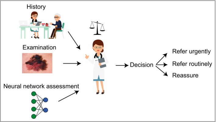
In the past, the skills required to make an accurate dermatological diagnosis have required exposure to thousands of patients over many years. However, in recent years, artificial intelligence (AI) has made enormous advances, particularly in the area of image classification. This has led computer scientists to apply these techniques to develop algorithms that are able to recognize skin lesions, particularly melanoma. Since 2017, there have been numerous studies assessing the accuracy of algorithms, with some reporting that the accuracy matches or surpasses that of a dermatologist. While the principles underlying these methods are relatively straightforward, it can be challenging for the practising dermatologist to make sense of a plethora of unfamiliar terms in this domain. Here we explain the concepts of AI, machine learning, neural networks and deep learning, and explore the principles of how these tasks are accomplished. We critically evaluate the studies that have assessed the efficacy of these methods and discuss limitations and potential ethical issues. The burden of skin cancer is growing within the Western world, with major implications for both population skin health and the provision of dermatology services. AI has the potential to assist in the diagnosis of skin lesions and may have particular value at the interface between primary and secondary care. The emerging technology represents an exciting opportunity for dermatologists, who are the individuals best informed to explore the utility of this powerful novel diagnostic tool, and facilitate its safe and ethical implementation within healthcare systems.


Figures

Figure 1
Figure 1
Schematic depicting how a machine learning algorithm trains on a large dataset to be able to match data to label (supervised learning), the performance of which can then be assessed.
Figure 2
Figure 2
Schematic of a receiver operating characteristic (ROC) curve, which is a way of visualizing the performance of a trained model's sensitivity and specificity. Typically, machine learning studies will use ROC curves and calculations of the area under the curve (AUC or AUROC) to quantify accuracy. The dashed line represents the desired perfect performance, when sensitivity and specificity are both 100%; in this scenario, the AUC would be 1·0. In reality, there is a trade‐off between sensitivity and specificity, which gives rise to a curve.
Figure 3
Figure 3
Schematic depicting how classification tasks are performed in convolutional neural networks. Pixel data from an image are passed through an architecture consisting of multiple layers of connecting nodes. In convolutional neural networks, these layers contain unique ‘convolutional layers’, which operate as filters. These filters work because it was recognized that the location of a feature within an image is often less important than whether that feature is present or absent – an example might be (theoretically) the presence or absence of blue‐grey veiling within a melanoma. A convolutional ‘filter’ learns a particular feature of the image irrespective of where it occurs within the image (represented by the black squares). The network is composed of a large number of hierarchical filters that learn increasingly high‐level representations of the image. These could in principle learn dermoscopic features similar to those described by clinicians, although in practice the precise features recognized are likely to differ from classic diagnostic criteria.
Figure 4
Figure 4
Receiver operating characteristic (ROC) curves from studies by Esteva et al.,14 Brinker et al.19, 20 and Tschandl et al.21 Most often, the dermatologists’ comparative ROC curves are plotted as individual data points. Lying below the curve means that their sensitivity and specificity, and therefore accuracy, are considered inferior to those of the model in the study. The studies all demonstrate that, on average, dermatologists sit below the ROC curve of the machine learning algorithm. It is noticeable that the performance of the clinicians in Brinker's studies (b, c), for example, is inferior to that of the clinicians in the Esteva study (a). Although there is a greater spread of clinical experience in the Brinker studies, the discrepancy could also be related to how the clinicians were tested. In both Brinker's and Tschandl's studies, some individual data points represent performance discrepancy that is significantly lower than data would suggest in the real world, which could suggest that the assessments may be biased against clinicians. AUC, area under the curve; CNN, convolutional neural network. All figures are reproduced with permission of the copyright holders.
Figure 5
Figure 5
Schematic showing hypothetical use of a machine learning algorithm to help nonexpert clinicians risk‐stratify lesions to make clinical decisions. Clinicians routinely weigh up both the benefits and limitations of common diagnostic aids such as prostate‐specific antigen or D‐dimers. Currently, there are very few useful dermatological diagnostic decision aids available to nonexpert clinicians, as the diagnostic process is dominated by image recognition. Convolutional neural network could represent a new class of decision aid that could help nonexpert clinicians triage appropriately and narrow down their differential diagnosis.

Comment in

References

    1. Turing AMI. Computing machinery and intelligence. Mind 1950; LIX:433–60.
    1. LeCun Y, Bengio Y, Hinton G. Deep learning. Nature 2015; 521:436–44. - PubMed
    1. LeCun Y, Boser BE, Denker JS et al Handwritten digit recognition with a back‐propagation network In: Advances in Neural Information Processing Systems 2 (Touretzky DS, ed.). Burlington, MA: Morgan‐Kaufmann, 1990; 396–404.
    1. Cireşan DC, Meier U, Gambardella LM, Schmidhuber J. Deep, big, simple neural nets for handwritten digit recognition. Neural Comput 2010; 22:3207–20. - PubMed
    1. Krizhevsky A, Sutskever I, Hinton GE. ImageNet classification with deep convolutional neural networks. Neural Inform Proc Systems 2012; 25:3065386.

Publication types