Effect of Therapy on Radiographic Progression in Axial Spondyloarthritis: A Systematic Review and Meta-Analysis
- PMID: 31960614
- PMCID: PMC7218689
- DOI: 10.1002/art.41206
Effect of Therapy on Radiographic Progression in Axial Spondyloarthritis: A Systematic Review and Meta-Analysis
Abstract
Objective: To investigate the effect of therapies on radiographic progression in patients with axial spondyloarthritis (SpA).
Methods: A comprehensive database search for studies assessing radiographic progression in axial SpA (particular treatment versus no treatment of interest) was performed. Study-specific standardized mean differences in treatment outcomes at 2 and ≥4 years were estimated and combined using random-effects models.
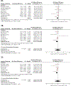
Results: Twenty-four studies in patients with axial SpA were identified, of which 18 involved tumor necrosis factor inhibitors (TNFi), 8 involved nonsteroidal antiinflammatory drugs (NSAIDs), and 1 involved secukinumab. Spinal radiographic progression, as measured by the modified Stoke Ankylosing Spondylitis Spine Score (mSASSS), was not significantly different between TNFi-treated and biologics-naive patients at 2 years (mSASSS difference -0.73 [95% confidence interval (95% CI) -1.52, 0.12], I2 = 28%) and ≥4 years (mSASSS difference -2.03 [95% CI -4.63, 0.72], I2 = 63%). Sensitivity analyses restricted to studies with a low risk of bias showed a significant difference in spinal radiographic progression between TNFi-treated and biologics-naive patients at ≥4 years (mSASSS difference -2.17 [95% CI -4.19, -0.15]). No significant difference in spinal radiographic progression was observed between NSAID-treated and control patients (mSASSS difference -0.30 [95% CI -2.62, 1.31], I2 = 71%) or between secukinumab-treated and biologics-naive patients (mSASSS difference -0.34 [95% CI -0.85, 0.17]). With regard to treatment differences in patients with nonradiographic axial SpA or in patients with radiographic progression measured using the sacroiliac joint score, an insufficient number of studies were available for analysis.
Conclusion: Although no significant protective effect of TNFi treatment on spinal radiographic progression was seen over the course of 2 years or ≥4 years in patients with axial SpA, our analysis restricted to studies with a low risk of bias showed a protective effect of TNFi after ≥4 years. Therefore, long-term TNFi exposure might confer beneficial effects on spinal radiographic progression in axial SpA. No difference in radiographic progression at 2 years was seen in either the NSAID or secukinumab treatment groups compared to their controls. Future studies should explore the effects of biologic treatment on radiographic progression, as well as the effects of long-term biologics exposure, in patients with early axial SpA or those with nonradiographic axial SpA.
© 2020, American College of Rheumatology.
Figures


Comment in
-
Challenges in Systematic Reviews That Include Observational Studies: Comment on the Article by Karmacharya et al.Arthritis Rheumatol. 2020 Dec;72(12):2163-2164. doi: 10.1002/art.41442. Arthritis Rheumatol. 2020. PMID: 32686909 No abstract available.
-
Reply.Arthritis Rheumatol. 2020 Dec;72(12):2164-2165. doi: 10.1002/art.41443. Epub 2020 Oct 3. Arthritis Rheumatol. 2020. PMID: 32686914 No abstract available.
References
-
- Ward MM, Deodhar A, Gensler LS, et al. 2019 Update of the American College of Rheumatology/Spondylitis Association of America/Spondyloarthritis Research and Treatment Network Recommendations for the Treatment of Ankylosing Spondylitis and Nonradiographic Axial Spondyloarthritis. Arthritis Rheumatol Hoboken NJ Published Online First: 22 August 2019. doi:10.1002/art.41042 - DOI - PMC - PubMed
-
- Braun J, Golder W, Bollow M, et al. Imaging and scoring in ankylosing spondylitis. Clin Exp Rheumatol 2002;20:S-178–S-184. - PubMed
-
- Oostveen J, Prevo R, den Boer J, et al. Early detection of sacroiliitis on magnetic resonance imaging and subsequent development of sacroiliitis on plain radiography. A prospective, longitudinal study. J Rheumatol 1999;26:1953–8. - PubMed
Publication types
MeSH terms
Grants and funding
LinkOut - more resources
Full Text Sources
Other Literature Sources
Research Materials

