. 2020 Jun 11;221(12):1978-1988.
doi: 10.1093/infdis/jiaa014.

Sensory Nociceptive Neurons Contribute to Host Protection During Enteric Infection With Citrobacter rodentium

Affiliations

Sensory Nociceptive Neurons Contribute to Host Protection During Enteric Infection With Citrobacter rodentium

Valerie T Ramirez et al. J Infect Dis. .

Abstract

Background: Neurons are an integral component of the immune system that functions to coordinate responses to bacterial pathogens. Sensory nociceptive neurons that can detect bacterial pathogens are found throughout the body with dense innervation of the intestinal tract.

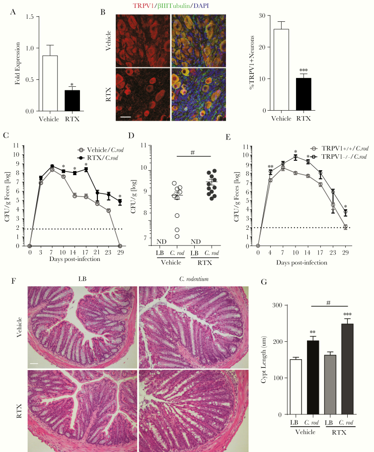
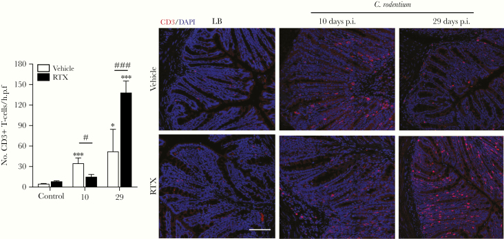
Methods: In this study, we assessed the role of these nerves in the coordination of host defenses to Citrobacter rodentium. Selective ablation of nociceptive neurons significantly increased bacterial burden 10 days postinfection and delayed pathogen clearance.

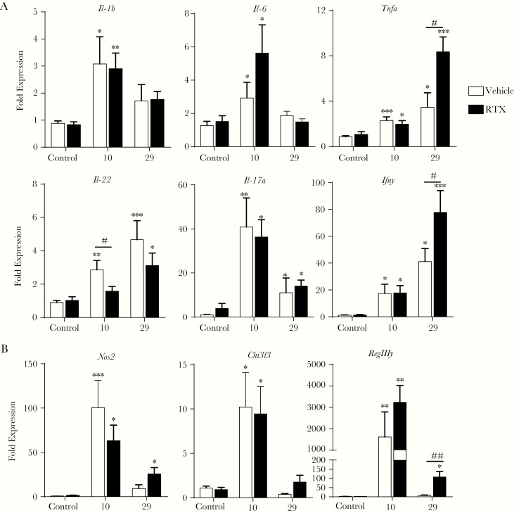
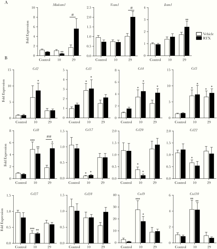
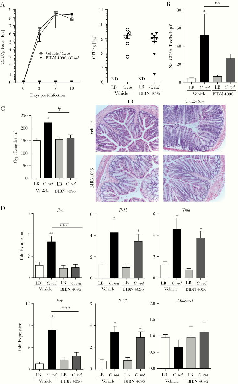
Results: Because the sensory neuropeptide CGRP (calcitonin gene-related peptide) regulates host responses during infection of the skin, lung, and small intestine, we assessed the role of CGRP receptor signaling during C rodentium infection. Although CGRP receptor blockade reduced certain proinflammatory gene expression, bacterial burden and Il-22 expression was unaffected.

Conclusions: Our data highlight that sensory nociceptive neurons exert a significant host protective role during C rodentium infection, independent of CGRP receptor signaling.

Keywords: Citrobacter rodentium; CGRP; TRPV1; nociceptors.


Figures

Figure 1.
Figure 1.
Ablation of nociceptive neurons increases the severity of infection and delays clearance of Citrobacter rodentium. Trpv1 message in the colon (10–12 mice/group) (A) and transient receptor potential cation channel subfamily V member 1 (TRPV1) immunoreactive neurons in the nodose ganglion (B) were assessed 20 days after administration of resiniferatoxin (RTX) or vehicle (5–6 mice/group; scale bar, 30 µm). Mice subjected to sensory nociceptive ablation were assessed for host response to C rodentium infection. The bacterial burden was assessed in the feces every 3–4 days over the course of the infection (C) and adherent to the colon at 10 days postinfection (p.i.) (15–16 animals/group, 3 independent experiments, dotted line: limit of detection) (D). TRPV1+/+ and TRPV1−/− were infected with C rodentium, and the bacterial burden in the feces were measured every 3–4 days (E). The effect of this infection on the colonic histopathology at 10 days p.i. (F and G) was determined by crypt length in ≥20 well oriented crypts per animal (7–9 mice/group, from 3 independent experiments). Scale bar, 50 µm. Data are presented as mean ± standard error of the mean: *, P < .05, **, P < .01, and ***, P < .0001 vs uninfected mice; #, P < .05 compared with vehicle C rodentium-infected mice; Student’s t test (A and B) and two-way analysis of variance (ANOVA) (C) and one-way ANOVA (D and G) with Tukey posttest. CFU, colony-forming units; DAPI, 4’,6-diamidino-2-phenylindole; LB, Luria-Bertani.
Figure 2.
Figure 2.
Reduced Il-22 expression in Citrobacter rodentium-infected mice lacking nociceptive neurons. The host immune response to C rodentium infection was determined by using quantitative polymerase chain reaction from mice with intact sensory nociceptor (vehicle) and ablated nociceptor (resiniferatoxin [RTX]) at 10 and 29 days postinfection. These included the expression of prototypical proinflammatory cytokines produced by innate and adaptive immune cells (A), M1 and M2 macrophages, and antimicrobial peptide (B) with 10–14 mice/group. Data are presented as mean ± standard error of the mean: *, P < .05, **, P < .01, and ***, P < .0001 versus uninfected mice; #, P < .05 and ##, P < .001 compared with vehicle C rodentium-infected mice; one-way analysis of variance with Tukey posttest.
Figure 3.
Figure 3.
Delayed T-cell recruitment during Citrobacter rodentium infection after nociceptor ablation. Colonic tissue sections were assessed for recruitment of T cells (CD3+ DAPI+) in vehicle ± C rodentium and resiniferatoxin (RTX) ± C rodentium-treated mice and quantified. Scale bar, 50 µm. Data are presented as mean ± standard error of the mean: *, P < .05 and ***, P < .001 compared with uninfected mice; #, P < .05 and ###, P < .001 compared with vehicle C rodentium-infected mice; one-way analysis of variance with Tukey posttest, with 8–12 animals per group. DAPI, 4’,6-diamidino-2-phenylindole; LB, Luria-Bertani; p.i., postinfection.
Figure 4.
Figure 4.
Loss of sensory innervation alters adhesion molecule expression in the colon during Citrobacter rodentium infection. Expression of adhesion molecules (A) and chemokines (B) involved in the homing and migration of adaptive immune cells into the colonic mucosa were evaluated by quantitative polymerase chain reaction at 10 and 29 days postinfection (p.i.). Data are presented as mean ± standard error of the mean: *, P < .05, **, P < .01, and ***, P < .001 versus uninfected mice; #, P < .05 and ##, P < .01 compared with vehicle C rodentium-infected mice; one-way analysis of variance with Tukey posttest, with 8–12 animals per group.
Figure 5.
Figure 5.
Calcitonin gene-related peptide (CGRP) receptor blockade alters select aspects of host immune function but does not affect Citrobacter rodentium bacterial burden. The role of CGRP signaling during C rodentium infection was assessed by administration of vehicle or BIBN 4096 starting on the day of the infection with consecutive injections every other day. The effect on host response was assessed by fecal bacterial burden and adherent bacterial burden at days 10 postinfection (A). T-cell recruitment, (B) colonic crypt hyperplasia, (C) scale bar, 50 µm, and (D) colonic expression of proinflammatory cytokines and Madcam1. Data are presented as mean ± standard error of the mean: *, P < .05 and **, P < .01 versus uninfected mice; #, P < .05 and ###, P < .001 compared with vehicle C rodentium-infected mice; one-way analysis of variance with Tukey posttest, with 6–10 animals per group. CFU, colony-forming units; LB, Luria-Bertani; ns, no significant difference.

References

    1. Reardon C, Murray K, Lomax AE. Neuroimmune communication in health and disease. Physiol Rev 2018; 98:2287–316. - PMC - PubMed
    1. Pinho-Ribeiro FA, Verri WA Jr, Chiu IM. Nociceptor sensory neuron-immune interactions in pain and inflammation. Trends Immunol 2017; 38:5–19. - PMC - PubMed
    1. Feng B, Gebhart GF. Characterization of silent afferents in the pelvic and splanchnic innervations of the mouse colorectum. Am J Physiol Gastrointest Liver Physiol 2011; 300:G170–80. - PMC - PubMed
    1. Brierley SM, Hibberd TJ, Spencer NJ. Spinal afferent innervation of the colon and rectum. Front Cell Neurosci 2018; 12:467. - PMC - PubMed
    1. Alawi K, Keeble J. The paradoxical role of the transient receptor potential vanilloid 1 receptor in inflammation. Pharmacol Ther 2010; 125:181–95. - PubMed

Publication types

MeSH terms

Substances