. 2020 Feb;26(2):255-265.
doi: 10.3201/eid2602.190346.

Porcine Deltacoronavirus Infection and Transmission in Poultry, United States1

Porcine Deltacoronavirus Infection and Transmission in Poultry, United States1

Patricia A Boley et al. Emerg Infect Dis. 2020 Feb.

Abstract

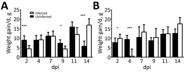
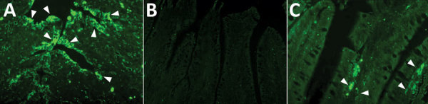
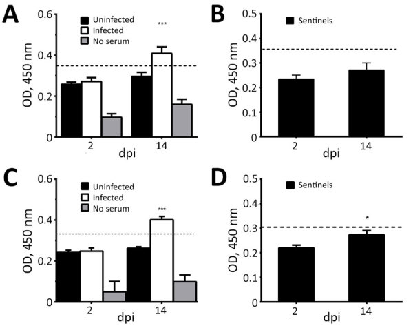
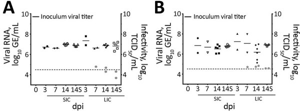
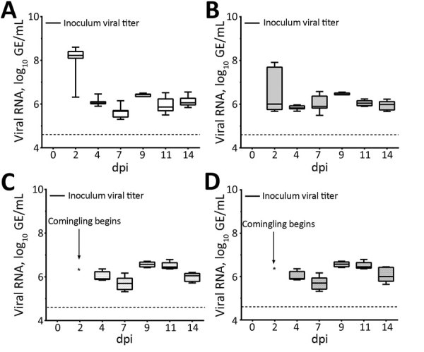
Coronaviruses cause respiratory and gastrointestinal diseases in diverse host species. Deltacoronaviruses (DCoVs) have been identified in various songbird species and in leopard cats in China. In 2009, porcine deltacoronavirus (PDCoV) was detected in fecal samples from pigs in Asia, but its etiologic role was not identified until 2014, when it caused major diarrhea outbreaks in swine in the United States. Studies have shown that PDCoV uses a conserved region of the aminopeptidase N protein to infect cell lines derived from multiple species, including humans, pigs, and chickens. Because PDCoV is a potential zoonotic pathogen, investigations of its prevalence in humans and its contribution to human disease continue. We report experimental PDCoV infection and subsequent transmission among poultry. In PDCoV-inoculated chicks and turkey poults, we observed diarrhea, persistent viral RNA titers from cloacal and tracheal samples, PDCoV-specific serum IgY antibody responses, and antigen-positive cells from intestines.

Keywords: Porcine deltacoronavirus; United States; chickens; coronaviruses; interspecies transmission; poultry; swine disease; turkeys; viruses; zoonoes.


Figures

Figure 1
Figure 1
Average weight gain/day of (A) chicks and (B) turkey poults in a study of infection and transmission of porcine deltacoronavirus in poultry. Weights were taken at 2, 4, 7, 9, 11, and 14 dpi and differences were averaged by the number of days between time points. Weights for sentinel birds are excluded after 2 dpi. Error bars indicate upper half of SD. Statistically significant values are indicated: *p<0.05; **p<0.01; ***p<0.001. dpi, days postinoculation.
Figure 2
Figure 2
Detection of porcine deltacoronavirus (PDCoV) antigen in poultry by immunofluorescent (IF) staining in the intestines of poults inoculated with intestinal contents of a PDCoV-infected GF pig (DC175) (6.87 log10 50% tissue culture infectious dose/mL) or a mock inoculate in a study of infection and transmission of porcine deltacoronavirus in poultry. A) PDCoV-infected pig intestine used as positive control; white arrows indicate widespread antibody staining. B) IF-stained jejunum of a poult (no. 42) at 14 dpi with no antigen-positive cells. C) IF-stained jejunum of a poult (no. 63) at 14 days postinoculation (dpi); white arrows indicate several PDCoV antigen-positive cells in the villous epithelial cells. Original magnification ×300.
Figure 3
Figure 3
Detection of porcine deltacoronavirus (PDCoV)–specific IgY antibody titers in serum collected from chicks and turkey poults in a study of infection and transmission of porcine deltacoronavirus in poultry. A) Uninfected (n = 6) and infected chicks (n = 6). B) Sentinel chicks (n = 5). C) Uninfected (n = 5) and infected poults (n = 6). D) Sentinel poults (n = 5). OD values are blanked against a control of uncoated wells (carbonate buffer only). Values represent the average of 3 replicates; error bars represent upper half of SEM. Dashed line indicates cutoff, which was determined by using the mean of uninfected birds at 2 dpi +2 SDs. Statistically significant values: *p<0.05; ***p<0.001. dpi, days postinoculation; OD, optical density.
Figure 4
Figure 4
Porcine deltacoronavirus (PDCoV) viral RNA shedding patterns in samples collected from cloaca (A) and trachea (B) of infected and uninfected chicks, and from cloaca (C) and trachea (D) of sentinel chicks in a study of infection and transmission of porcine deltacoronavirus in poultry. Inoculum viral titer represents the genomic equivalent (GE) of inoculum administered at onset, 9.71 log10 GE/mL. Boxplots represent distribution of values; tops and bottoms of boxes represent 10%–90% range of values; horizontal lines within boxes indicate medians; error bars represent SEM. Dashed line indicates detection limit of 4.6 log10 GE/mL of PDCoV in samples. dpi, days postinoculation; GE, genomic equivalent.
Figure 5
Figure 5
Viral RNA titers and infectivity of intestinal contents of (A) chicks and (B) poults in a study of infection and transmission of porcine deltacoronavirus in poultry. Inoculum viral titer represents the genomic equivalent (GE) of inoculum administered at onset, 9.71 log10 GE/mL. Gray dots represent infectivity at 7 and 14 dpi, expressed in log10 TCID50/mL, as indicated on the right y-axis. Dashed line indicates detection limit of 4.6 log10 GE/mL of PDCoV in samples. Shapes represent individual birds necropsied at each time point. Solid bars represent the mean. dpi, days postinoculation; GE, genomic equivalent; LIC, large intestine contents; S, sentinel birds necropsied; SIC, small intestine contents; TCID50, 50% tissue culture infectious dose.
Figure 6
Figure 6
Porcine deltacoronavirus (PDCoV) viral RNA shedding patterns in samples collected from cloaca (A) and trachea (B) of infected and uninfected poults and from cloaca (C) and trachea (D) of sentinel poults in a study of infection and transmission of porcine deltacoronavirus in poultry. Inoculum viral titer represents the genomic equivalent (GE) of inoculum administered at onset, 9.71 log10 GE/mL. Box plots represent distribution of values; tops and bottoms of boxes represent 10%–90% range of values; horizontal lines within boxes indicate medians; error bars represent SEM. Dashed line indicates detection limit of 4.6 log10 GE/mL of PDCoV in samples. dpi, days postinoculation.

References

    1. Masters PS, Perlman S. Coronaviridae. In: Knipe DM, Howley PM, eds. Fields virology, 6th ed. Philadelphia: Lippincott Williams & Wilkins; 2013. p. 825–58.
    1. Chan JF, To KK, Tse H, Jin DY, Yuen KY. Interspecies transmission and emergence of novel viruses: lessons from bats and birds. Trends Microbiol. 2013;21:544–55. 10.1016/j.tim.2013.05.005 - DOI - PMC - PubMed
    1. Schwegmann-Wessels C, Herrler G. Transmissible gastroenteritis virus infection: a vanishing specter. Dtsch Tierarztl Wochenschr. 2006;113:157–9. - PubMed
    1. Song D, Zhou X, Peng Q, Chen Y, Zhang F, Huang T, et al. Newly emerged porcine deltacoronavirus associated with diarrhoea in swine in China: identification, prevalence and full-length genome sequence analysis. Transbound Emerg Dis. 2015;62:575–80. 10.1111/tbed.12399 - DOI - PMC - PubMed
    1. Huang YW, Dickerman AW, Piñeyro P, Li L, Fang L, Kiehne R, et al. Origin, evolution, and genotyping of emergent porcine epidemic diarrhea virus strains in the United States. MBio. 2013;4:e00737–13. 10.1128/mBio.00737-13 - DOI - PMC - PubMed

Publication types

LinkOut - more resources