. 2020 Jul;68(7):1361-1374.
doi: 10.1002/glia.23783. Epub 2020 Jan 21.

Astrocyte-T cell crosstalk regulates region-specific neuroinflammation

Affiliations

Astrocyte-T cell crosstalk regulates region-specific neuroinflammation

Jessica L Williams et al. Glia. 2020 Jul.

Abstract

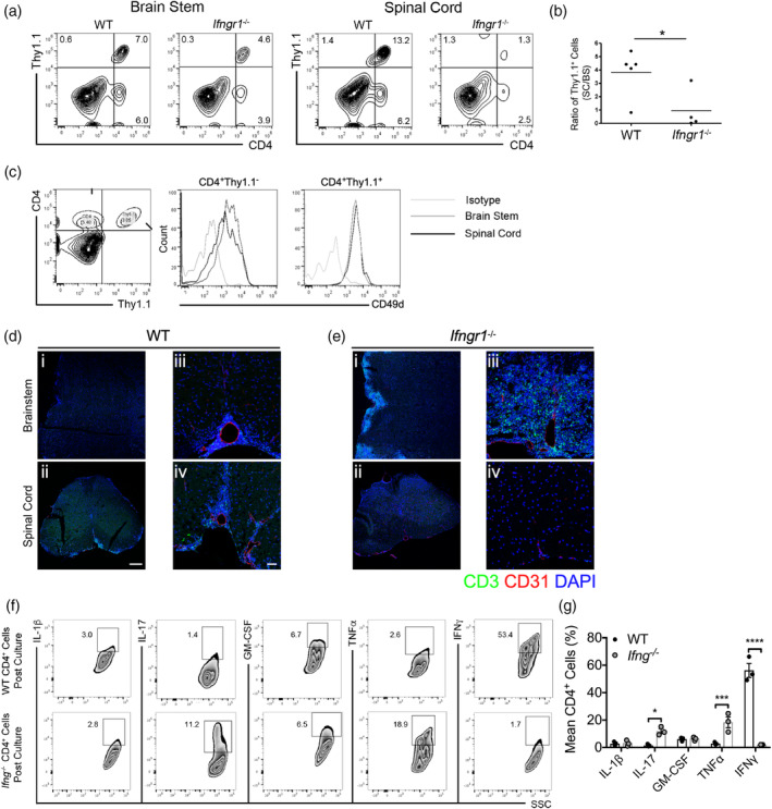
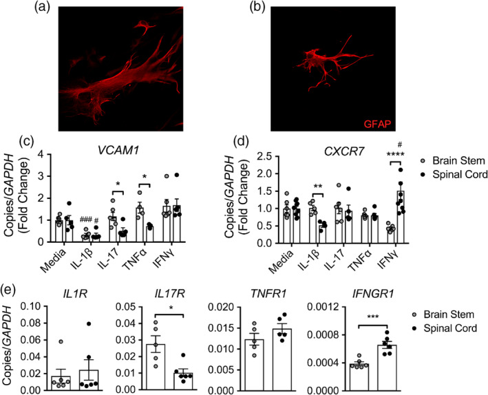
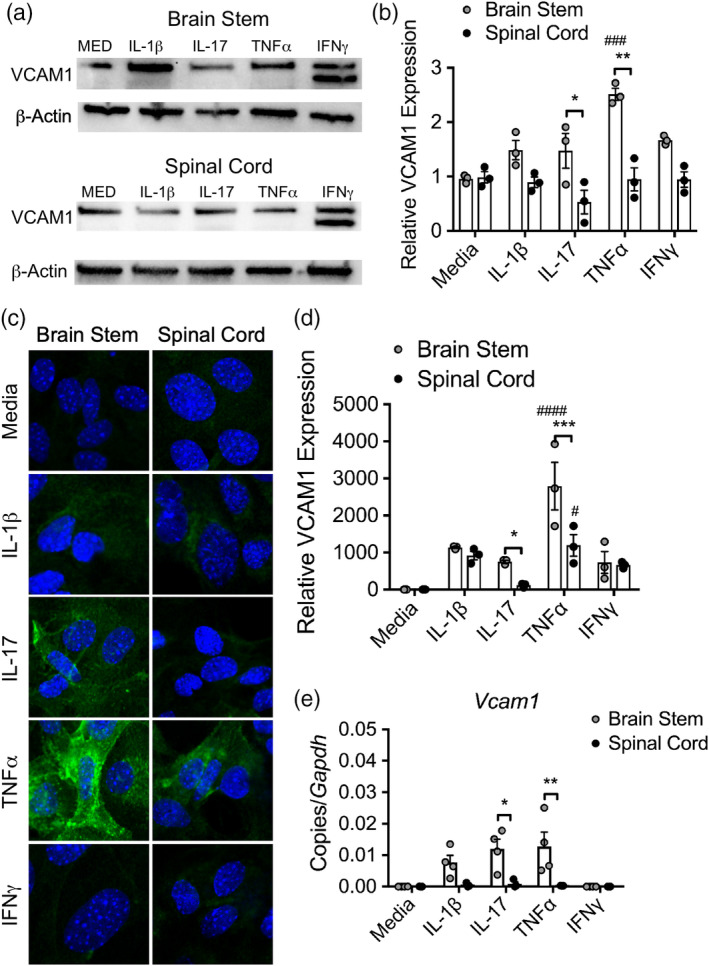
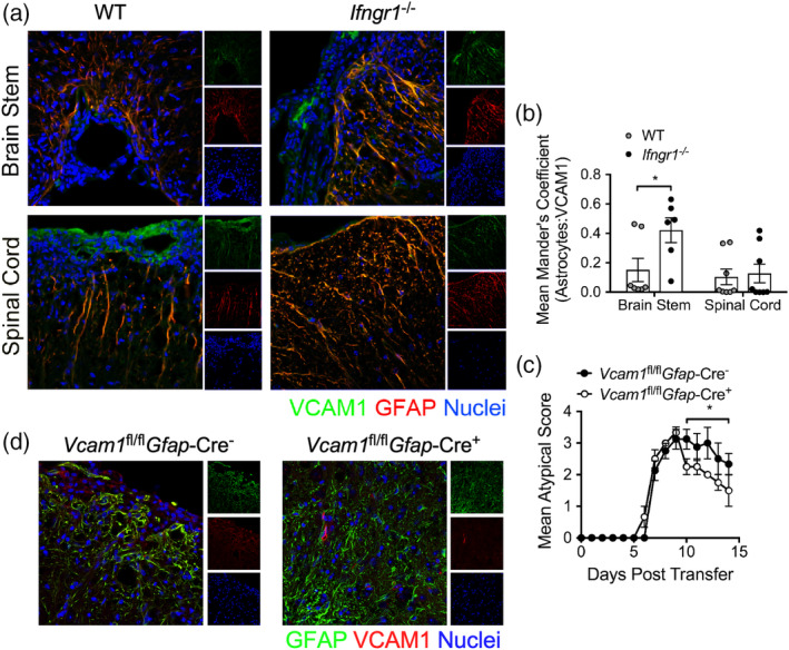
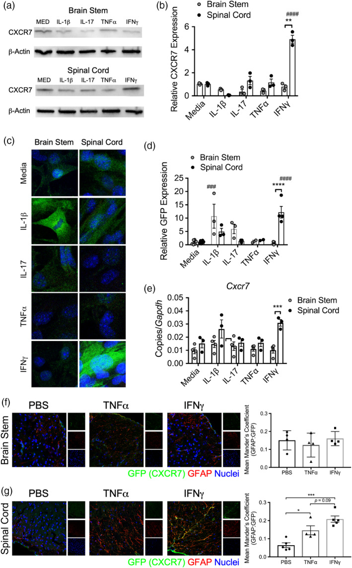
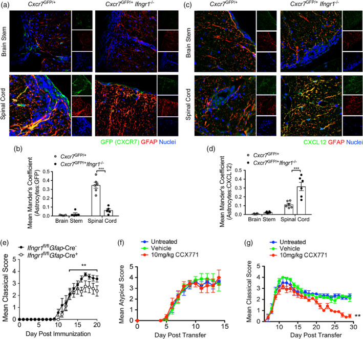
During multiple sclerosis (MS), an inflammatory and neurodegenerative disease of the central nervous system (CNS), symptoms, and outcomes are determined by the location of inflammatory lesions. While we and others have shown that T cell cytokines differentially regulate leukocyte entry into perivascular spaces and regional parenchymal localization in murine models of MS, the molecular mechanisms of this latter process are poorly understood. Here, we demonstrate that astrocytes exhibit region-specific responses to T cell cytokines that promote hindbrain versus spinal cord neuroinflammation. Analysis of cytokine receptor expression in human astrocytes showed region-specific responsiveness to Th1 and Th17 inflammatory cytokines. Consistent with this, human and murine astrocytes treated with these cytokines exhibit differential expression of the T cell localizing molecules VCAM-1 and CXCR7 that is both cytokine and CNS region-specific. Using in vivo models of spinal cord versus brain stem trafficking of myelin-specific T cells and astrocyte-specific deletion strategies, we confirmed that Th1 and Th17 cytokines differentially regulate astrocyte expression of VCAM-1 and CXCR7 in these locations. Finally, stereotaxic injection of individual cytokines into the hindbrain or spinal cord revealed region- and cytokine-specific modulation of localizing cue expression by astrocytes. These findings identify a role for inflammatory cytokines in mediating local astrocyte-dependent mechanisms of immune cell trafficking within the CNS during neuroinflammation.

Keywords: CXCR7; T cell; VCAM-1; astrocyte; cytokine; neuroinflammation; regional heterogeneity.


Conflict of interest statement

The authors declare no competing financial interests.

Figures

Figure 1
Figure 1
Cytokines dictate regional T cell trafficking within the CNS. (a) Activated, myelin‐specific Thy1.1+ T cell clones were injected into WT or Ifngr1 −/− recipients. CNS leukocytes were collected during peak disease (10 days post‐transfer) and the presence of donor and recipient T cells was analyzed. (b) The number of spinal cord Thy1.1+ cells was calculated and normalized to the number of Thy1.1+ cells isolated from the brain stem. (c) The level of CD49d on donor and recipient CD4+ T cells isolated from the brain stem (gray line) and spinal cord (black line) was analyzed and compared to an isotype control (light gray line) by flow cytometry. (d) Activated, myelin‐specific WT T cells were injected into naïve WT and (e) Ifngr1 −/− recipients and at peak EAE. CNS tissue was cryopreserved then labeled for the expression of CD3 and CD31. A representative, (i and ii) tiled 20× magnification image; scale bar = 200 μm, and (iii and iv) a 20× image are shown; scale bar = 50 μm. (f) Leukocytes were isolated from the draining lymph nodes of immunized WT or Ifng −/− mice and restimulated in the presence of MOG35‐55 for 72 hr. Following restimulation, intracellular cytokine expression in CD4+ cells was analyzed by flow cytometry. (g) Cytokine expression in CD4+ cells was quantified over three independent experiments. *p < .05; ***p < .001, ****p < .0001 by Student's t test or two‐way ANOVA. CNS, central nervous system; EAE, experimental autoimmune encephalomyelitis
Figure 2
Figure 2
Cytokines mediate regional localization cues on astrocytes. Primary human astrocytes from the (a) brain stem and (b) spinal cord were labeled for GFAP by ICC and imaged using confocal microscopy at 20× magnification. (c) CXCR7 and (d) VCAM1 mRNA levels in primary adult human brain stem and spinal cord astrocytes following exposure to 10 ng/ml of recombinant cytokine for 24 hr. (e) Cytokine receptor expression in primary adult human brain stem and spinal cord astrocytes was quantified by qRT‐PCR following culture in astrocyte medium. Data shown are combined results from two independent experiments with n = 5–8 per treatment. *p < .05; **p < .01; ***p < .001, ****p < .0001 between region and # p < .05; ### p < .001 compared to media‐treated astrocytes by Student's t test or two‐way ANOVA. ICC, immunocytochemistry
Figure 3
Figure 3
Brain stem astrocytes preferentially upregulate VCAM‐1 in response to Th17‐associated cytokines. (a) WB analysis of lysate from human brain stem and spinal cord astrocytes following exposure to 10 ng/ml cytokine for 24 hr was probed for VCAM‐1. (b) VCAM‐1 density was quantified and normalized to media controls. (c) Primary murine astrocytes from the brain stem and spinal cord of neonatal pups were exposed to 10 ng/ml of recombinant cytokine for 24 hr, fixed and analyzed for VCAM‐1 expression by confocal microscopy. (d) The level of expression was quantified using the AxioVision image analysis software and normalized to media‐treated astrocytes. (e) Following cytokine exposure, mRNA transcript was isolated from murine astrocytes and Vcam1 expression was measured by qRT‐PCR. Data shown are combined results from three to four independent experiments. *p < .05; **p < .01; ***p < .001 between region and # p < .05; ### p < .001 compared to media‐treated astrocytes by two‐way ANOVA. WB, western blot
Figure 4
Figure 4
Brain stem inflammation is maintained by VCAM‐1 on astrocytes during EAE. (a) Activated WT, myelin‐specific Th1 clones were injected into WT or Ifngr1 −/− mice and the CNS was prepared for IHC at peak disease. The brain stem and spinal cord of recipient mice were labeled for VCAM‐1 and GFAP expression. (b) The mean Mander's coefficient for colocalization (ImageJ) was used to quantify astrocyte‐associated VCAM‐1 expression in each CNS region. Data are representative of two independent experiments and data points represent individual animals. (c) Activated, myelin‐specific Ifng −/− T cells were injected into Vcam1 fl/fl Gfap‐Cre or Vcam1 fl/fl Gfap‐Cre+ littermates and monitored for signs of atypical EAE. (d) Representative 20× confocal images of brain stem tissue from Vcam1 fl/fl Gfap‐Cre and Vcam1 fl/fl Gfap‐Cre+ littermates labeled for IHC to confirm reduced VCAM‐1 expression in GFAP+ astrocytes. *p < .05 by two‐way ANOVA or Mann–Whitney U test for EAE clinical scores. CNS, central nervous system; EAE, experimental autoimmune encephalomyelitis; IHC, immunohistochemistry
Figure 5
Figure 5
Spinal cord astrocytes express CXCR7 in response to IFNγ. (a) WB analysis of lysate from human brain stem and spinal cord astrocytes following exposure to 10 ng/ml cytokine for 24 hr was probed for CXCR7. (b) CXCR7 density was quantified and normalized to media controls. (c) Primary murine astrocytes from the brain stem and spinal cord of neonatal Cxcr7 GFP/+ pups were exposed to 10 ng/ml of recombinant cytokine for 24 hr, fixed and analyzed for GFP expression by confocal microscopy. (d) The level of GFP expression was quantified using the AxioVision image analysis software and normalized to media‐treated astrocytes. (e) Following cytokine exposure, mRNA transcript was isolated from murine astrocytes and Cxcr7 expression was measured by qRT‐PCR. Data shown are combined results from three to four independent experiments. About 25 ng of recombinant mouse TNFα, IFNγ, or vehicle was injected into the brain stem or spinal cord of Cxcr7 GFP/+ mice. About 72 hr following injection, brain stem and spinal cord sections were harvested and prepared for IHC. (e) Brain stem and (f) spinal cord sections were stained and analyzed for expression of GFP and GFAP. The mean Mander's coefficient for colocalization (ImageJ) was used to quantify astrocyte‐associated GFP. Data are pooled from two independent experiments per CNS region. *p < .05; ***p < .001 by two‐way ANOVA. CNS, central nervous system; IHC, immunohistochemistry; WB, western blot
Figure 6
Figure 6
Astrocyte IFNγ signaling regulates the CXCR7/CXCL12 chemokine axis to maintain spinal cord inflammation. (a–d) Activated WT, myelin‐specific Th1 clones were injected into either Cxcr7 GFP/+ or Cxcr7 GFP/+ Ifngr1 −/− mice and the CNS was prepared for IHC at peak disease. The brain stem and spinal cord of recipient mice were labeled for GFAP and (a) GFP or (c) CXCL12. The mean Mander's coefficient for colocalization (ImageJ) was used to quantify astrocyte‐associated (b) GFP and (d) CXCL12 expression in each CNS region. Data are representative of two independent experiments. (e) Ifngr1 fl/fl Gfap‐Cre or Ifngr1 fl/fl Gfap‐Cre+ littermates were immunized with MOG35‐55 and adjuvants and monitored for signs of classical EAE. Data are representative of three independent experiments with n = 5–8 mice per group. Activated (f) WT or (g) Ifng −/− myelin‐specific T cells were injected into WT mice. At the onset of EAE, mice were either untreated or subcutaneously injected with 100 μl captisol or 10 mg/kg CCX771 in 100 μl captisol daily. (f) Classical or (g) atypical signs of EAE were monitored. Data are representative of two to three independent experiments with n = 4–7 mice per group. ***p < .001, by two‐way ANOVA and **p < .01 by Mann–Whitney U test for EAE clinical scores. CNS, central nervous system; EAE, experimental autoimmune encephalomyelitis; IHC, immunohistochemistry

References

    1. Abbott, N. J. , Patabendige, A. A. , Dolman, D. E. , Yusof, S. R. , & Begley, D. J. (2010). Structure and function of the blood–brain barrier. Neurobiology of Disease, 37(1), 13–25. 10.1016/j.nbd.2009.07.030 - DOI - PubMed
    1. Arellano, G. , Ottum, P. A. , Reyes, L. I. , Burgos, P. I. , & Naves, R. (2015). Stage‐specific role of interferon‐gamma in experimental autoimmune encephalomyelitis and multiple sclerosis. Frontiers in Immunology, 6, 492 10.3389/fimmu.2015.00492 - DOI - PMC - PubMed
    1. Bever, C. T., Jr. , Panitch, H. S. , Levy, H. B. , McFarlin, D. E. , & Johnson, K. P. (1991). Gamma‐interferon induction in patients with chronic progressive MS. Neurology, 41(7), 1124–1127. - PubMed
    1. Billiau, A. , Heremans, H. , Vandekerckhove, F. , Dijkmans, R. , Sobis, H. , Meulepas, E. , & Carton, H. (1988). Enhancement of experimental allergic encephalomyelitis in mice by antibodies against IFN‐gamma. Journal of Immunology, 140(5), 1506–1510. - PubMed
    1. Booth, H. D. E. , Hirst, W. D. , & Wade‐Martins, R. (2017). The role of astrocyte dysfunction in Parkinson's disease pathogenesis. Trends in Neurosciences, 40(6), 358–370. 10.1016/j.tins.2017.04.001 - DOI - PMC - PubMed

Publication types

MeSH terms