. 2020 Feb;53(2):e12720.
doi: 10.1111/cpr.12720. Epub 2020 Jan 21.

Circular RNA hsa_circ_0061825 (circ-TFF1) contributes to breast cancer progression through targeting miR-326/TFF1 signalling

Affiliations

Circular RNA hsa_circ_0061825 (circ-TFF1) contributes to breast cancer progression through targeting miR-326/TFF1 signalling

Gaofeng Pan et al. Cell Prolif. 2020 Feb.

Abstract

Objectives: Circular RNAs (circRNAs) are RNA transcripts that belong to non-coding RNAs (ncRNAs), whose implication in human cancers has been recently demonstrated. However, the specific role of multiple circRNAs in breast cancer remains unidentified.

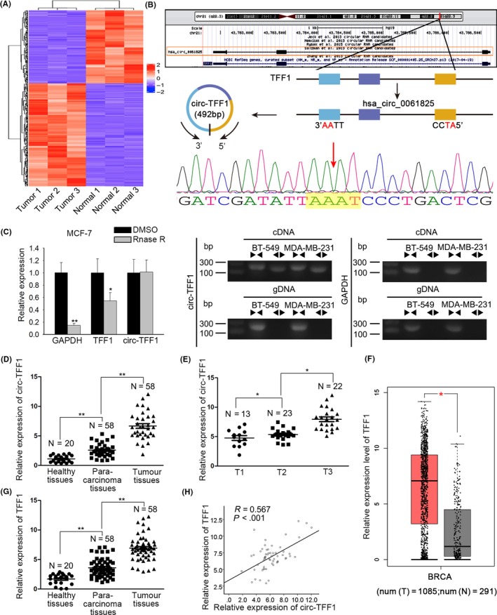
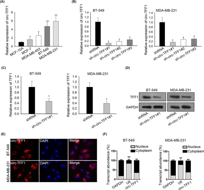
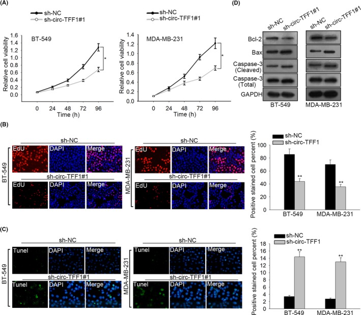
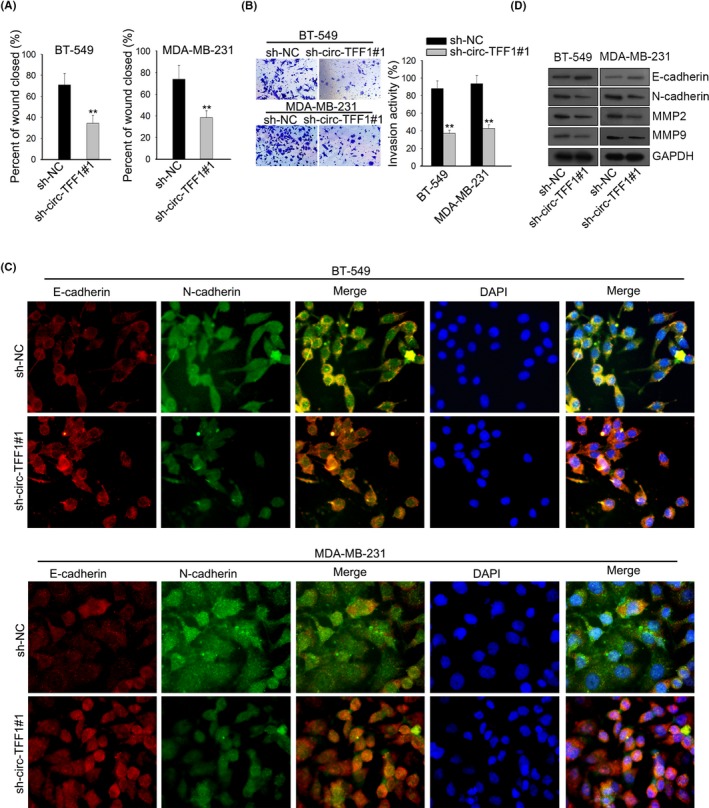
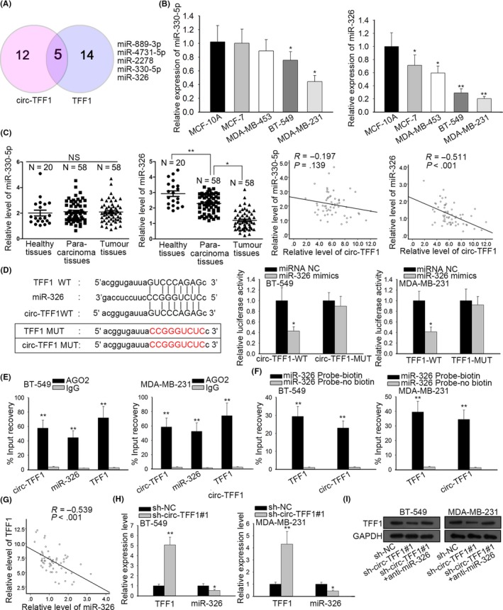
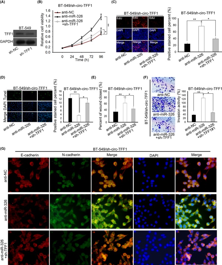
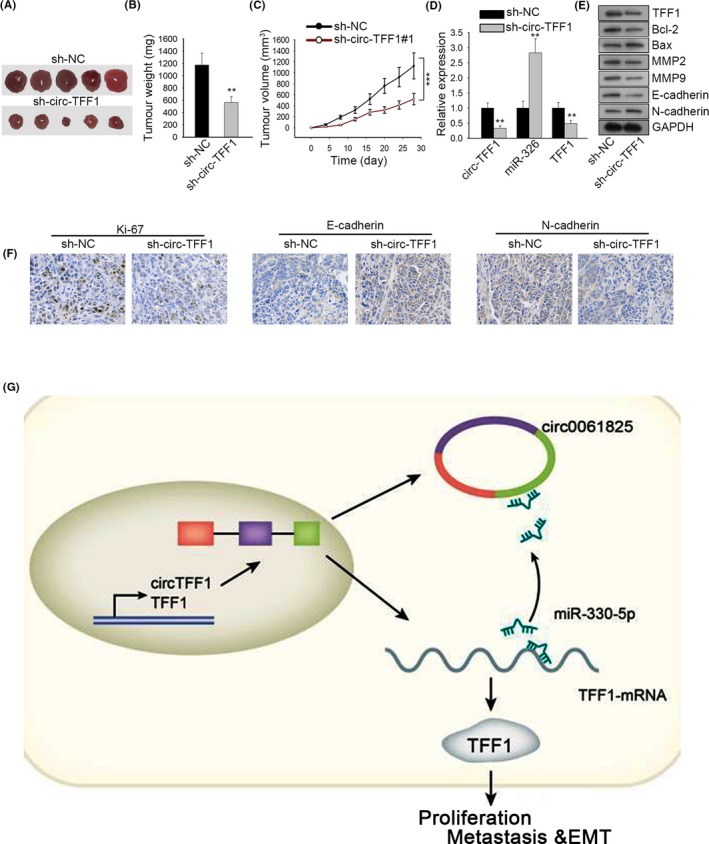
Materials and methods: Microarray analysis and bioinformatics analysis were applied to select circRNA and miRNA, respectively. The loop structure of circ-TFF1 was confirmed using RNase R treatment, divergent primer PCR and Sanger sequencing. qRT-PCR and Western blot were employed for gene expressions. In vitro and in vivo experiments were conducted to assess the function of circ-TFF1 in biological processes in breast cancer cells. FISH and subcellular separation indicated circ-TFF1 cellular distribution. Luciferase reporter and RIP assays and Pearson's correlation analysis were performed to evaluate relationships between genes.

Results: Circ-TFF1 and TFF1 were both upregulated and positively associated with each other in breast cancer. Knockdown of circ-TFF1 hindered breast cancer cell proliferation, migration, invasion and EMT in vitro and controlled tumour growth in vivo. Circ-TFF1 acted as a ceRNA of TFF1 by sponging miR-326, and its contribution to breast cancer progression was mediated by miR-326/TFF1 axis.

Conclusions: Circ-TFF1 is a facilitator in breast cancer relying on TFF1 by absorbing miR-326, providing a novel promising target for BC treatment.

Keywords: TFF1; breast cancer; circ-TFF1; miR-326.


Conflict of interest statement

The authors indicate no conflicts of interest in this study.

Figures

Figure 1
Figure 1
Hsa_circ_0061825 (circ‐TFF1) and TFF1 were both upregulated in breast cancer and in positive association with each other. A, High‐throughput sequencing of circRNAs in tumour and normal tissues. B, The position of circ‐TFF1 in chromosome. C, The circular structure of circ‐TFF1 was verified by RNase R treatment, divergent primer PCR and Sanger sequencing. D‐E, qRT‐PCR results of circ‐TFF1 expression in healthy, para‐carcinoma and tumour tissues and in T1, T2 and T3 stages. F, The high expression of TFF1 in breast carcinoma tissues was gained by TCGA database. G, qRT‐PCR was used for detecting TFF1 expression in healthy, tumorous and non‐tumour tissues. H, Pearson's correlation analysis was utilized for the association between circ‐TFF1 and TFF1. *P < .05, **P < .01
Figure 2
Figure 2
circ‐TFF1 modulated TFF1 positively. A, In breast cancer cells including MDA‐MB‐231, BT‐549, MCF‐7 and MDA‐MB‐453 and normal breast epithelial cells MCF‐10A, circ‐TFF1 expression was examined by qRT‐PCR. B, The transfection efficiency of sh‐circ‐TFF1#1/2/3 in BT‐549 and MDA‐MB‐231 cells was confirmed through qRT‐PCR. (C‐D) TFF1 expression affected by sh‐circ‐TFF1#1 was evaluated by qRT‐PCR and Western blotting. E‐F, Assays of FISH and qRT‐PCR after subcellular fractionation were adopted for the location of circ‐TFF1 in breast cancer cells. *P < .05, **P < .01
Figure 3
Figure 3
Silencing of circ‐TFF1 inhibited the proliferation and induced the apoptosis in breast cancer. A‐B, CCK‐8 and EdU analyses of cell proliferation in BT‐549 and MDA‐MB‐231 cells transfected with sh‐NC or sh‐circ‐TFF1#1. C, Tunel analysis of cell apoptosis affected by sh‐circ‐TFF1#1 in BT‐549 and MDA‐MB‐231 cells. D, Levels of cell apoptosis‐related proteins (Bcl2, Bax, caspase3 (cleaved), total‐caspase3) were assessed through Western blot. *P < .05, **P < .01
Figure 4
Figure 4
Silencing of circ‐TFF1 repressed metastasis and EMT process of breast cancer. A‐B, The migration and invasion abilities of two cells were separately estimated through would healing assay and transwell assay. C, Levels of N‐cadherin and E‐cadherin by sh‐circ‐TFF1#1 in two cells were detected via IF assay. D, Western blot was performed to test EMT‐related proteins involving N‐cadherin, E‐cadherin, MMP2 and MMP9. **P < .01
Figure 5
Figure 5
Circ‐TFF1 targets miR‐326 to promote TFF1 expression. A, Five miRNAs shared by circ‐TFF1 and TFF1 were acquired from starBase. The Venn diagram was exhibited. B‐C, The respective levels of miR‐330‐5p and miR‐326 in breast cancer cells and breast epithelial cells, as well as in healthy, para‐carcinoma and tumour tissues were analysed by qRT‐PCR. And the relationship between circ‐TFF1 and miR‐330‐5p or miR‐326 was dissected with Pearson's correlation analysis. D, The wild‐type and mutant binding sites of circ‐TFF1 or TFF1 for miR‐326 were constructed. The interaction between miR‐326 and circ‐TFF1 or TFF1 was confirmed by luciferase reporter assay in 293T cells. E‐F, The relationship among circ‐TFF1, miR‐326 and TFF1 was validated using RIP and RNA pull‐down assays. G, Pearson's correlation analysis was utilized for exploring the association of miR‐326 with TFF1. H, qRT‐PCR detection of the impact of sh‐circ‐TFF1#1 on TFF1 and miR‐326. I, Western blot results of TFF1 expression under transfection of sh‐circ‐TFF1#1 or co‐transfection of sh‐circ‐TFF1#1 and miR‐326 inhibitor. *P < .05, **P < .01
Figure 6
Figure 6
TFF1 downregulation abolished the miR‐326 inhibition‐promoted cellular processes in circ‐TFF1‐silenced BT‐549 cells. A, The inference efficacy of sh‐TFF1#1 was testified by Western blotting. B‐C, Cell proliferation of three indicated groups was individually tested by CCK‐8 and EdU assays. D, Tunel assay was carried out for detecting cell apoptosis in different groups. E‐F, Wound healing and transwell experiments were conducted to estimate cell motility in three groups. G, IF analysis of E‐cadherin and N‐cadherin levels of different groups. *P < .05, **P < .01
Figure 7
Figure 7
Circ‐TFF1 promotes tumour formation in vivo. A, Images of tumours in sh‐circ‐TFF1#1 or sh‐NC group. B‐C, The difference of tumour weight and the growth curve of tumour volume in two groups were portrayed. D, Expression of circ‐TFF1, miR‐326 and TFF1 in tumours derived from circ‐TFF1‐inhibited BT‐549 cells was determined by qRT‐PCR. E, Western blotting results of the levels of TFF1, Ki67, Bcl‐2, Bax, MMP2, MMP9, E‐cadherin and N‐cadherin when circ‐TFF1 was silenced. F, IHC staining of Ki67, E‐cadherin and N‐cadherin in the paraffin‐embedded sections of tumours collected above. G, The final regulatory circuit of circ‐TFF1‐miR‐326‐TFF1 axis in breast cancer progression. **P < .01, ***P < .001

References

    1. Torre LA, Bray F, Siegel RL, Ferlay J, Lortet‐Tieulent J, Jemal A. Global cancer statistics, 2012. CA Cancer J Clin. 2015;65(2):87‐108. - PubMed
    1. Mahvi DA, Liu R, Grinstaff MW, Colson YL, Raut CP. Local cancer recurrence: The realities, challenges, and opportunities for new therapies. CA Cancer J Clin. 2018. - PMC - PubMed
    1. DeSantis C, Ma J, Bryan L, Jemal A. Breast cancer statistics, 2013. CA Cancer J Clin. 2014;64(1):52‐62. - PubMed
    1. Danan M, Schwartz S, Edelheit S, Sorek R. Transcriptome‐wide discovery of circular RNAs in Archaea. Nucleic Acids Res. 2012;40(7):3131‐3142. - PMC - PubMed
    1. Jeck WR, Sharpless NE. Detecting and characterizing circular RNAs. Nat Biotechnol. 2014;32(5):453‐461. - PMC - PubMed