. 2020 Mar;9(5):1768-1778.
doi: 10.1002/cam4.2748. Epub 2020 Jan 21.

Breast cancer stromal clotting activation (Tissue Factor and thrombin): A pre-invasive phenomena that is prognostic in invasion

Affiliations

Breast cancer stromal clotting activation (Tissue Factor and thrombin): A pre-invasive phenomena that is prognostic in invasion

Hudhaifah Shaker et al. Cancer Med. 2020 Mar.

Abstract

Background: Tumor stroma, of which fibroblasts are the most abundant cell, resembles a non-healing wound, where a procoagulant environment creates a permissive milieu for cancer growth. We aimed to determine if tumor expression of coagulation factors (procoagulant phenotype), and systemic hypercoagulability, occur at the preinvasive (ductal carcinoma in situ; DCIS) stage and correlate with breast cancer subtype, disease-free survival (DFS), and overall survival (OS).

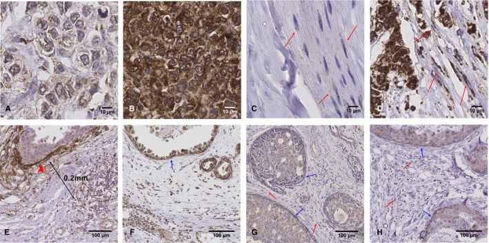
Methods: In a prospective cohort of early breast cancer (DCIS, n = 76; invasive, n = 248) tumor, normal breast and plasma were examined. Fibroblast and epithelial expression of Tissue Factor (TF), thrombin, PAR1, PAR2, and plasma thrombin-antithrombin (TAT) and D-dimer were correlated with clinicopathological data, and 5-year survival.

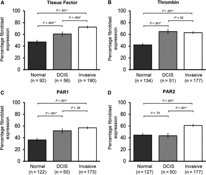
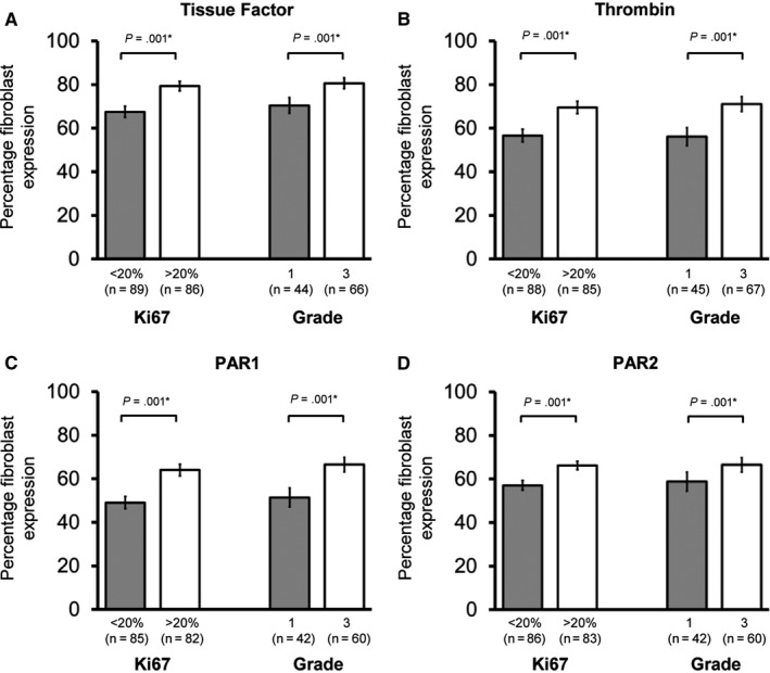
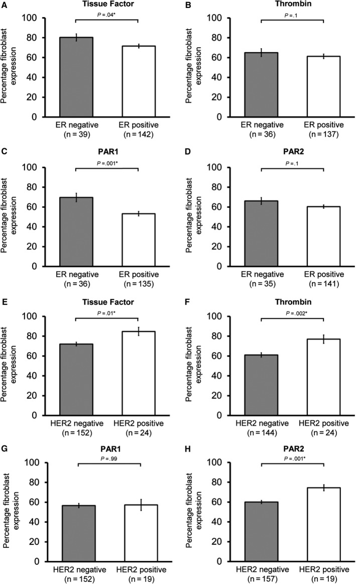
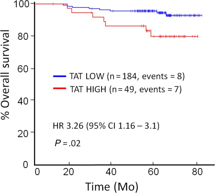
Results: Fibroblast expression of TF, thrombin, and PAR1 was increased in DCIS and invasive cancer compared to normal breast fibroblasts (P ≤ .003, all). Fibroblast TF, thrombin, PAR1, and PAR2 was increased in cancers with high Ki67, high grade, ER- (vs ER+), and HER2+ (vs HER2-) (all P < .05). On univariate analysis, fibroblast TF expression was inversely associated with DFS (P = .04) and OS (P = .02). D-dimer was higher in node positive (507 (CI: 411-625) ng/mL, n = 68) vs negative patients (428 (CI: 387-472) ng/mL, n = 171, P = .004) and inversely associated with OS (P = .047). On multivariate analysis, plasma TAT was associated with reduced OS (HR 3.26, CI 1.16-3.1, P = .02), with a high plasma TAT (≥3.2 ng/mL) associated with > 3-fold mortality risk compared to low TAT.

Conclusion: This demonstrates procoagulant phenotypic changes occur in fibroblasts at the preinvasive stage. Fibroblast procoagulant phenotype is associated with aggressive breast cancer subtypes and reduced survival. Coagulation may be a therapeutic target in breast cancer.

Keywords: DCIS; PAR1; PAR2; breast cancer; coagulation; fibroblast; thrombin; thrombosis; tissue factor.


Conflict of interest statement

The authors declare no potential conflicts of interest.

Figures

Figure 1
Figure 1
Tissue Microarray scoring of epithelial and stromal fibroblast extrinsic clotting pathway expression. Row 1: Thrombin epithelial staining: Images of invasive breast cancer tissue Immunohistochemistry (IHC)‐stained for thrombin with (A) low and (B) high epithelial staining. Row 1: Thrombin stromal staining and fibroblast scoring: Images of tissue IHC‐stained for thrombin with (C) <20% of stromal fibroblasts exhibiting staining and (D) 80% of stromal fibroblasts exhibiting staining. Example fibroblasts are pointed out with red arrows. Row 2: Tissue Factor (TF), thrombin, PAR1, and PAR2 ductal carcinoma in situ (DCIS) fibroblast scoring. DCIS tissue IHC stained for (E) Tissue Factor (F) thrombin (G) PAR1 (H) PAR2. In general, TF demonstrated strong myoepithelial staining (arrowhead, figure E). To avoid the potential risk of overscoring fibroblast TF staining, through including myoepithelial cells, TF fibroblast scoring was performed at a minimum of 0.2 mm away from the duct (Figure 1E). Intense myoepithelial staining was not a feature of thrombin, PAR1 or PAR2 IHC stains. (Blue arrows: myoepithelial cells, red arrows: fibroblasts)
Figure 2
Figure 2
Stromal expression of the extrinsic clotting pathway is increased in ductal carcinoma in situ (DCIS) and invasive breast cancer. Fibroblast expression of (A) Tissue Factor, (B) thrombin, (C) PAR1, and (D) PAR2 in the CHAMPion cohort is shown. Data is presented as mean percentage fibroblasts with positive expression ± Standard error of the mean (SEM). Number of samples tested shown in brackets. Statistical differences between groups were tested using ANOVA and Fisher's Least Significant Differences test for post hoc analyses. Normal, normal breast tissue; DCIS, ductal carcinoma in situ tissue; Invasive, invasive breast cancer tissue
Figure 3
Figure 3
Fibroblast expression of the extrinsic clotting pathway is increased in high proliferation and high‐grade invasive breast cancers. Fibroblast expression of (A) Tissue Factor, (B) thrombin, (C) PAR1, and (D) PAR2 according to Ki67 expression and grade (1 vs 3) in invasive cancer patients is shown. Data are presented as mean percentage fibroblasts with positive expression ± Standard error of the mean (SEM). Number of samples tested shown in brackets. Statistical differences between groups were tested using Student's t test
Figure 4
Figure 4
Fibroblast expression of the extrinsic clotting pathway is increased in estrogen receptor (ER)‐negative and HER2‐positive breast cancer. Fibroblast expression of (A) Tissue Factor, (B) thrombin, (C) PAR1, and (D) PAR2 in ER‐negative and ER‐positive cancers and (E) Tissue Factor, (F) thrombin, and (G) PAR2 in HER2‐negative and HER2‐positive cancers is shown. Data are presented as mean percentage fibroblasts with positive expression ± Standard error of the mean (SEM). Number of samples tested shown in brackets. Statistical differences between groups were tested using Student's t test. ER, oestrogen receptor. HER2, Human epidermal growth factor receptor 2
Figure 5
Figure 5
Kaplan‐Meier survival curves for overall survival according to plasma thrombin‐antithrombin (TAT). The preoperative plasma TAT was dichotomised into high (≥3.2 ng/mL) and low (<3.2 ng/mL). Association between plasma TAT and overall survival is shown. Univariate statistical testing was performed using Cox proportional hazards model (multivariate testing is detailed in Appendix D). Multivariate p‐value and hazard ratio shown

References

    1. World Cancer Research Fund. https://www.wcrf.org/dietandcancer/cancer-trends/breast-cancer-statistics. Accessed April, 2019.
    1. Sharma M, Beck AH, Webster JA, et al. Analysis of stromal signatures in the tumor microenvironment of ductal carcinoma in situ. Breast Cancer Res Treat. 2010;123(2):397‐404. - PMC - PubMed
    1. Dumont N, Liu B, Defilippis RA, et al. Breast fibroblasts modulate early dissemination, tumorigenesis, and metastasis through alteration of extracellular matrix characteristics. Neoplasia. 2013;15(3):249‐262. - PMC - PubMed
    1. Ostman A, Augsten M. Cancer‐associated fibroblasts and tumor growth–bystanders turning into key players. Curr Opin Genet Dev. 2009;19(1):67‐73. - PubMed
    1. Surowiak P, Murawa D, Materna V, et al. Occurence of stromal myofibroblasts in the invasive ductal breast cancer tissue is an unfavourable prognostic factor. Anticancer Res. 2007;27(4C):2917‐2924. - PubMed

Publication types

MeSH terms