Actin-like protein 8 promotes cell proliferation, colony-formation, proangiogenesis, migration and invasion in lung adenocarcinoma cells
- PMID: 31962007
- PMCID: PMC7049497
- DOI: 10.1111/1759-7714.13247
Actin-like protein 8 promotes cell proliferation, colony-formation, proangiogenesis, migration and invasion in lung adenocarcinoma cells
Abstract
Background: Non-small cell lung cancer (NSCLC) is the leading cause of cancer-associated mortality worldwide of which lung adenocarcinoma (LUAD) is the most common. The identification of oncogenes and effective drug targets is the key to individualized LUAD treatment. Actin-like protein 8 (ACTL8), a member of the cancer/testis antigen family, is associated with tumor growth and patient prognosis in various types of cancer. However, whether ACTL8 is involved in the development of LUAD remains unknown. The aim of the present study was to demonstrate the role of ACTL8 in human LUAD cells.
Methods: The expression of ACTL8 in LUAD tissues and cell lines was assessed using immunohistochemistry and western blotting. Additionally, plasmids expressing ACTL8-specific short hairpin RNAs were used to generate lentiviruses which were subsequently used to infect A549 and NCI-H1975 human LUAD cells. Cell proliferation, migration, invasion and apoptosis, as well as cell cycle progression and the expression of protein markers of epithelial to mesenchymal transition were investigated. A549 cell tumor growth in nude mice was also examined.
Results: The results showed that ACTL8 was highly expressed in A549 and NCI-H1975 LUAD cell lines. Additionally, ACTL8-knockdown inhibited proliferation, colony formation, cell cycle progression, migration and invasion, and increased apoptosis in both cell lines. Furthermore, in vivo experiments in nude mice revealed that ACTL8-knockdown inhibited A549 cell tumor growth.
Conclusion: These results suggest that ACTL8 serves an oncogenic role in human LUAD cells, and that ACTL8 may represent a potential therapeutic target for LUAD.
Key points: Our results suggest that ACTL8 serves an oncogenic role in human LUAD cells, and that ACTL8 may represent a potential therapeutic target for LUAD.
Keywords: ACTL8; angiogenesis; cell proliferation; lung adenocarcinoma; migration.
© 2019 The Authors. Thoracic Cancer published by China Lung Oncology Group and John Wiley & Sons Australia, Ltd.
Figures


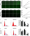
 ) shCtrl and (
 ) shACTL8.
 ) shCtrl and (
 ) shACTL8. (c) Flow cytometric analysis of A549 and NCI‐H1975 cells (d) three days after infection; the number of cells in the G0/G1 phase was increased, the number in the S phase was decreased, and the number in the G2/M phase was increased in the lenti‐shACTL8 group, compared with the control group. (
 ) shCtrl and (
 ) shACTL8. The data are presented as the mean ± standard deviation; n = 3. *P < 0.05 and **P < 0.01 versus lenti‐shCtrl. ACTL8, Actin‐like protein 8; LUAD, lung adenocarcinoma; sh, short‐hairpin; lenti, lentivirus; Ctrl, control.
 ) shCtrl and (
 ) shACTL8. (b) Apoptotic A549 and NCI‐H1975 cells were assessed using a TUNEL assay (magnification, 200x). (c) The expression of apoptosis‐related proteins apoptosis regulator BAX and Caspase 3 was determined by western blotting, and GAPDH was used as the loading control. ACTL8, Actin‐like protein 8; LUAD, lung adenocarcinoma; sh, short‐hairpin; lenti, lentivirus; Ctrl, control.
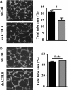
 ) shCtrl and (
 ) shACTL8.
 ) shCtrl and (
 ) shACTL8.
 ) shCtrl and (
 ) shACTL8.

 ) shCtrl and (
 ) shACTL8. (d) H&E staining of the excised tumors (magnification, x200). ACTL8, Actin‐like protein 8; sh, short‐hairpin; lenti, lentivirus; Ctrl, control.
Similar articles
-
USF1-induced overexpression of long noncoding RNA WDFY3-AS2 promotes lung adenocarcinoma progression via targeting miR-491-5p/ZNF703 axis.Mol Carcinog. 2020 Aug;59(8):875-885. doi: 10.1002/mc.23181. Epub 2020 Apr 10. Mol Carcinog. 2020. PMID: 32275336
-
Overexpression of KIF18A promotes cell proliferation, inhibits apoptosis, and independently predicts unfavorable prognosis in lung adenocarcinoma.IUBMB Life. 2019 Jul;71(7):942-955. doi: 10.1002/iub.2030. Epub 2019 Feb 28. IUBMB Life. 2019. PMID: 30817091
-
LncRNA LINC00680 promotes lung adenocarcinoma growth via binding to GATA6 and canceling GATA6-mediated suppression of SOX12 expression.Exp Cell Res. 2021 Aug 1;405(1):112653. doi: 10.1016/j.yexcr.2021.112653. Epub 2021 May 21. Exp Cell Res. 2021. PMID: 34029572
-
RBM15 facilities lung adenocarcinoma cell progression by regulating RASSF8 stability through N6 Methyladenosine modification.Transl Oncol. 2024 Aug;46:102018. doi: 10.1016/j.tranon.2024.102018. Epub 2024 Jun 4. Transl Oncol. 2024. PMID: 38838436 Free PMC article. Review.
-
Significance of stratifin in early progression of lung adenocarcinoma and its potential therapeutic relevance.Pathol Int. 2021 Oct;71(10):655-665. doi: 10.1111/pin.13147. Epub 2021 Jul 29. Pathol Int. 2021. PMID: 34324245 Review.
Cited by
-
[Lung Cancer Stem-like Cells and Drug Resistance].Zhongguo Fei Ai Za Zhi. 2022 Feb 20;25(2):111-117. doi: 10.3779/j.issn.1009-3419.2022.102.02. Zhongguo Fei Ai Za Zhi. 2022. PMID: 35224964 Free PMC article. Review. Chinese.
-
Therapeutic Effects of Perilla Phenols in Oral Squamous Cell Carcinoma.Int J Mol Sci. 2023 Oct 5;24(19):14931. doi: 10.3390/ijms241914931. Int J Mol Sci. 2023. PMID: 37834377 Free PMC article.
-
ACTL8 Promotes the Progression of Gastric Cancer Through PI3K/AKT/mTOR Signaling Pathway.Dig Dis Sci. 2024 Oct;69(10):3786-3798. doi: 10.1007/s10620-024-08649-6. Epub 2024 Sep 25. Dig Dis Sci. 2024. PMID: 39322809 Free PMC article.
-
Cancer/Testis Antigens as Biomarker and Target for the Diagnosis, Prognosis, and Therapy of Lung Cancer.Front Oncol. 2022 Apr 27;12:864159. doi: 10.3389/fonc.2022.864159. eCollection 2022. Front Oncol. 2022. PMID: 35574342 Free PMC article. Review.
-
Actin-Like Protein 8 Promotes the Progression of Triple-Negative Breast Cancer via Activating PI3K/AKT/mTOR Pathway.Onco Targets Ther. 2021 Apr 12;14:2463-2473. doi: 10.2147/OTT.S291403. eCollection 2021. Onco Targets Ther. 2021. PMID: 33883901 Free PMC article.
References
-
- Siegel RL, Miller KD, Jemal A. Cancer statistics, 2019. CA Cancer J Clin 2019; 69: 7–34. - PubMed
-
- Cafarotti S, Lococo F, Froesh P, Zappa F, Andre D. Target therapy in lung cancer. Adv Exp Med Biol 2016; 893: 127–36. - PubMed
-
- Casaluce F, Sgambato A, Maione P et al ALK inhibitors: A new targeted therapy in the treatment of advanced NSCLC. Target Oncol 2013; 8: 55–67. - PubMed
-
- Singh M, Jadhav HR. Targeting non‐small cell lung cancer with small‐molecule EGFR tyrosine kinase inhibitors. Drug Discov Today 2018; 23: 745–53. - PubMed
MeSH terms
Substances
LinkOut - more resources
Full Text Sources
Medical
Molecular Biology Databases

