. 2020 May;62(5):633-644.
doi: 10.1165/rcmb.2019-0092OC.

Impaired Myofibroblast Dedifferentiation Contributes to Nonresolving Fibrosis in Aging

Affiliations

Impaired Myofibroblast Dedifferentiation Contributes to Nonresolving Fibrosis in Aging

Kosuke Kato et al. Am J Respir Cell Mol Biol. 2020 May.

Abstract

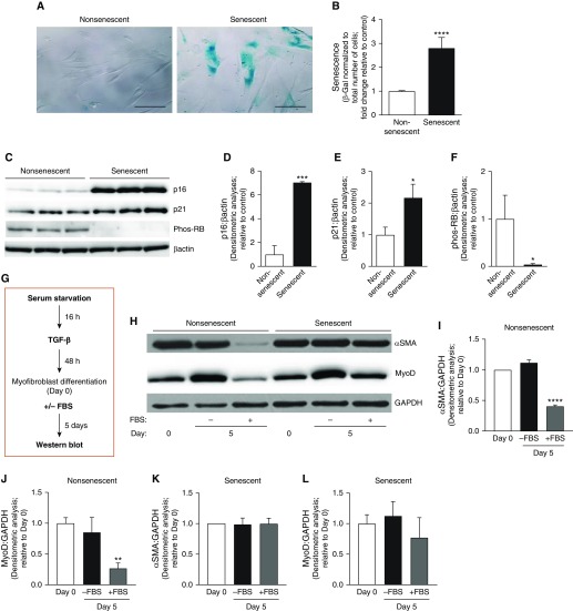
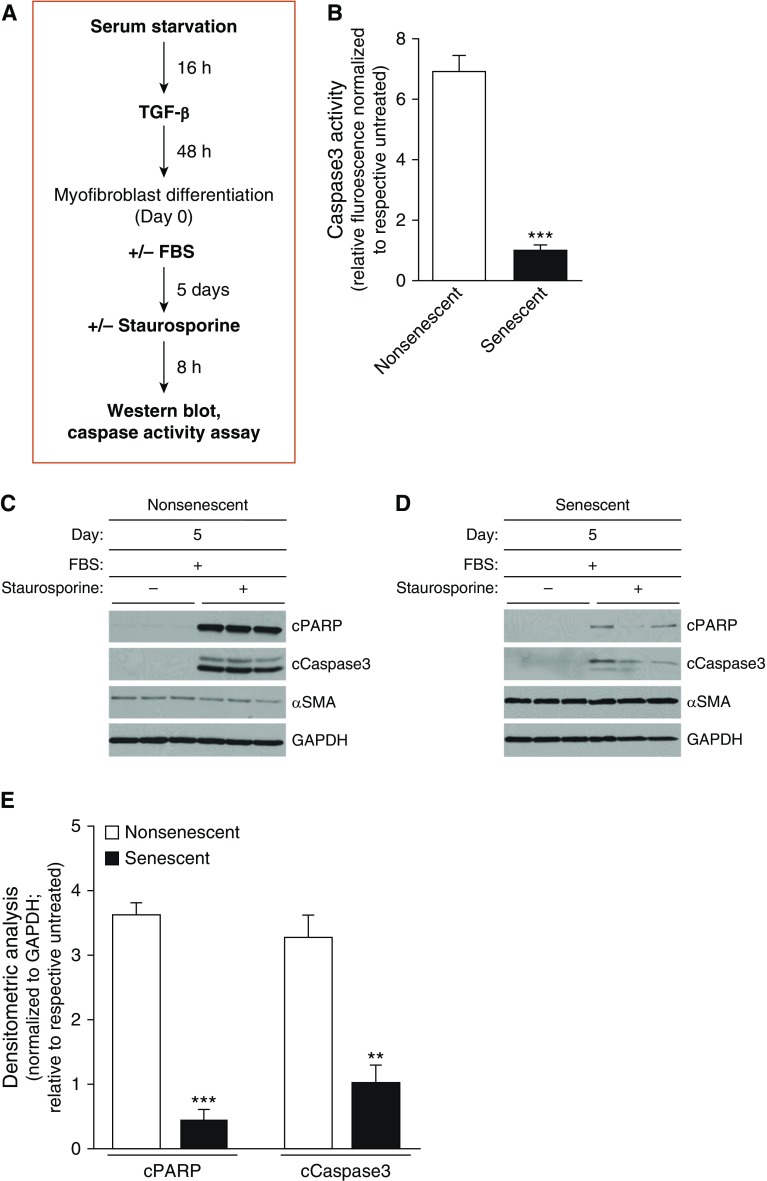
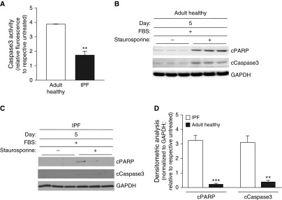
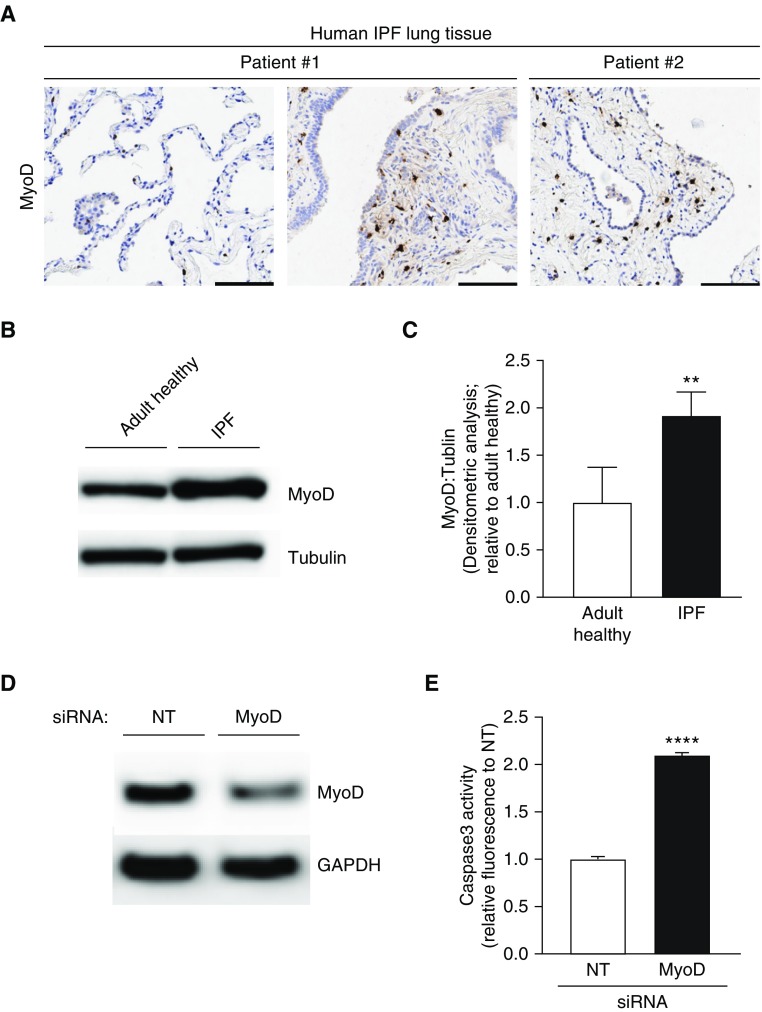
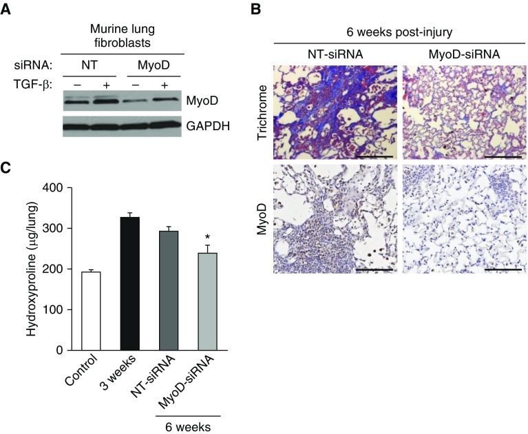
Idiopathic pulmonary fibrosis (IPF) is a fatal age-associated disease with no cure. Although IPF is widely regarded as a disease of aging, the cellular mechanisms that contribute to this age-associated predilection remain elusive. In this study, we sought to evaluate the consequences of senescence on myofibroblast cell fate and fibrotic responses to lung injury in the context of aging. We demonstrated that nonsenescent lung myofibroblasts maintained the capacity for dedifferentiation, whereas senescent/IPF myofibroblasts exhibited an impaired capacity for dedifferentiation. We previously demonstrated that the transcription factor MyoD acts as a critical switch in the differentiation and dedifferentiation of myofibroblasts. Here, we demonstrate that decreased levels of MyoD preceded myofibroblast dedifferentiation and apoptosis susceptibility in nonsenescent cells, whereas MyoD expression remained elevated in senescent/IPF myofibroblasts, which failed to undergo dedifferentiation and demonstrated resistance to apoptosis. Genetic strategies to silence MyoD restored the susceptibility of IPF myofibroblasts to undergo apoptosis and led to a partial reversal of age-associated persistent fibrosis in vivo. The capacity for myofibroblast dedifferentiation and subsequent apoptosis may be critical for normal physiologic responses to tissue injury, whereas restricted dedifferentiation and apoptosis resistance in senescent cells may underlie the progressive nature of age-associated human fibrotic disorders. These studies support the concept that senescence may promote profibrotic effects via impaired myofibroblast dedifferentiation and apoptosis resistance, which contributes to myofibroblast accumulation and ultimately persistent fibrosis in aging.

Keywords: MyoD; apoptosis resistance; myofibroblast plasticity; pulmonary fibrosis; senescence.


Figures

Figure 1.
Figure 1.
Senescent myofibroblasts have a diminished capacity for dedifferentiation. Lung fibroblasts at low and high population doublings (nonsenescent and senescent, respectively) were used and cultured ex vivo. (AF) Senescence was evaluated by qualitative (A) and quantitative (B) measurements of senescence-associated β-galactosidase (β-gal) activity, expression of the senescence markers by Western blotting (C), and densitometric analyses (DF). Scale bars: 100 μm. *P < 0.05, ***P < 0.001, and ****P < 0.0001 as compared with nonsenescent, using Student’s two-tailed t-test. (GL) Cells were serum starved for 16 hours and treated with TGF-β (transforming growth factor β; 2 ng/ml) for 48 hours. At 48 hours after TGF-β treatment (Day 0), the media was replaced with TGF-β–free media with 0% or 20% FBS and cells were incubated for 5 days. (G) Schematic diagram illustrating the treatment protocol. (H–L) Protein expression of α-SMA (α-smooth muscle actin), MyoD (myogenic differentiation), and GAPDH was assessed by Western blotting (H) and quantified by densitometric analyses (IL). **P < 0.01 and ****P < 0.0001 as compared with Day 0, using Student’s two-tailed t-test. All values represent means ± SEM; n = 3–5 biological replicates, 3–5 independent experiments. Phos-RB = phosphorylated retinoblastoma protein.
Figure 2.
Figure 2.
Idiopathic pulmonary fibrosis (IPF) lung myofibroblasts demonstrate impaired dedifferentiation capacity. (AD) Fibroblasts isolated from the lungs of healthy adults and patients with biopsy-proven IPF were evaluated for cellular senescence by qualitative (A) and quantitative (B) measurements of senescence-associated β-gal activity, expression of the senescence markers by Western blotting (C), and densitometric analyses (D). Scale bars: 100 μm. *P < 0.05 and ****P < 0.0001 as compared with adult healthy lungs, using Student’s two-tailed t-test. (EJ) Cells were serum starved for 16 hours, treated with TGF-β (2 ng/ml) for 48 hours (Day 0), and then treated with 0% or 20% FBS for 5 days. Expression of α-SMA, MyoD, and GAPDH was assessed by Western blotting in fibroblasts isolated from adult healthy lungs (E) and from IPF lungs (H). (F–J) α-SMA and MyoD were quantified by densitometric analyses for adult healthy fibroblasts (F and G) and for IPF fibroblasts (I and J). **P < 0.01 and ****P < 0.0001 as compared with Day 0, using Student’s two-tailed t-test. All values represent means ± SEM; n = 3 biological replicates from 3 independent experiments.
Figure 3.
Figure 3.
Impaired capacity for dedifferentiation in senescent myofibroblasts is associated with apoptosis resistance. Nonsenescent and senescent lung fibroblasts were serum starved for 16 hours, treated with TGF-β (2 ng/ml) for 48 hours (Day 0), and then treated with 0% or 20% FBS for 5 days. Cells were treated with vehicle or staurosporine (300 nM), an apoptosis-inducing agent, for 8 hours. (A) Schematic diagram illustrating the treatment protocol and endpoints assessed. (B) Caspase 3 activity was assessed. (CE) Expression of cPARP, cCaspase 3, α-SMA, and GAPDH was assessed by Western blotting (C and D) and by densitometric analyses (E). All values represent means ± SEM; n = 3 biological replicates from 2 independent experiments; **P < 0.01 and ***P < 0.001 as compared with nonsenescent, using Student’s two-tailed t-test. cPARP = cleaved poly-ADP-ribose polymerases.
Figure 4.
Figure 4.
Impaired dedifferentiation in IPF fibroblasts is associated with apoptosis resistance. Adult healthy and IPF lung fibroblasts were serum starved for 16 hours, treated with TGF-β (2 ng/ml) for 48 hours (Day 0), and then treated with 0% or 20% FBS for 5 days. Cells were treated with vehicle or staurosporine (300 nM) for 8 hours, and Caspase 3 activity was assessed (A). (BD) Expression of cPARP, cCaspase 3, and GAPDH was assessed by Western blotting (B and C) and densitometric analyses (D). Values represent means ± SEM; n = 3 biological replicates; **P < 0.01 and ***P < 0.001 using Student’s two-tailed t test.
Figure 5.
Figure 5.
MyoD is upregulated in IPF lung myofibroblasts, and genetic targeting restores apoptosis susceptibility. (A) IPF lung tissue sections were analyzed by immunohistochemistry for expression of MyoD. Scale bars: 100 μm. (B and C) Adult healthy and IPF lung fibroblasts were evaluated for protein expression of MyoD by Western blotting (B) and densitometric analyses (C). (D and E) IPF fibroblasts were transfected with MyoD siRNA. Downregulation of MyoD was confirmed by Western blotting (D) and caspase activity of transfected cells was assessed (E). Values represent means ± SEM; n = 3 biological replicates; **P < 0.01 and ****P < 0.0001 using Student’s two-tailed t test. NT = nontargeting siRNA.
Figure 6.
Figure 6.
MyoD is upregulated in the lungs of aged mice with persistent fibrosis. Young (2 mo old) and aged (18 mo old) C57BL/6 mice were subjected to lung injury by airway instillation of intratracheal bleomycin (1.25 U/kg). Lung tissue was harvested at 0 (uninjured), 3 weeks, and 2 months after injury. (A) Immunohistochemistry analysis of MyoD. Scale bars: 100 μm. (B) Densitometric analyses of MyoD protein expression in whole-lung tissues at 2 months after injury. Values represent means ± SEM; n = 4–5 biological replicates; *P < 0.05 compared with young mice using Student’s two-tailed t-test.
Figure 7.
Figure 7.
Therapeutic targeting of MyoD in the lungs of aged mice with established fibrosis leads to fibrosis resolution. (A) Isolated lung fibroblasts from uninjured young mice were transfected with MyoD-targeting or -nontargeting siRNA. Transfected cells were serum starved overnight and treated with/without TGF-β (2 ng/ml) for 48 hours. MyoD was assessed by Western blotting. (B and C) Aged (18 mo old) C57BL/6 mice were subjected to lung injury by airway instillation of intratracheal bleomycin (1.25 U/kg). The mice were then treated with intranasal instillation of MyoD or vehicle siRNA every other day from Week 3 to Week 6. Lung tissue was harvested at 0 week (uninjured), 3 weeks, and 6 weeks after injury/treatment. Fibrosis was assessed by Masson’s trichrome blue staining for collagen (top panels) and MyoD was assessed by immunohistochemical staining for MyoD (bottom panels) (B), and whole-lung homogenates were analyzed by quantitative hydroxyproline assay (C). Scale bars: 100 μm. Data are expressed as total micrograms of hydroxyproline per whole lung. Differences among groups were assessed with one-way ANOVA multiple comparisons with Tukey’s post test. Values represent means ± SEM; n = 6–11 biological replicates; *P < 0.05 as compared with NT-siRNA using one-way ANOVA multiple comparisons with Tukey’s post test.

Comment in

  • Senescence, the Janus of Lung Injury and Repair.
    Wallach-Dayan SB, Rojas M. Wallach-Dayan SB, et al. Am J Respir Cell Mol Biol. 2020 May;62(5):548-549. doi: 10.1165/rcmb.2020-0022ED. Am J Respir Cell Mol Biol. 2020. PMID: 31978311 Free PMC article. No abstract available.

References

    1. Wynn TA. Fibrotic disease and the T(H)1/T(H)2 paradigm. Nat Rev Immunol. 2004;4:583–594. - PMC - PubMed
    1. Wynn TA. Common and unique mechanisms regulate fibrosis in various fibroproliferative diseases. J Clin Invest. 2007;117:524–529. - PMC - PubMed
    1. Fell CD, Martinez FJ, Liu LX, Murray S, Han MK, Kazerooni EA, et al. Clinical predictors of a diagnosis of idiopathic pulmonary fibrosis. Am J Respir Crit Care Med. 2010;181:832–837. - PMC - PubMed
    1. Hecker L, Thannickal VJ. Nonresolving fibrotic disorders: idiopathic pulmonary fibrosis as a paradigm of impaired tissue regeneration. Am J Med Sci. 2011;341:431–434. - PubMed
    1. Collard HR. The age of idiopathic pulmonary fibrosis. Am J Respir Crit Care Med. 2010;181:771–772. - PubMed

Publication types