Review
. 2020 Jan 16;6(1):15.
doi: 10.3390/jof6010015.

Oral Candidiasis: A Disease of Opportunity

Affiliations
Review

Oral Candidiasis: A Disease of Opportunity

Taissa Vila et al. J Fungi (Basel). .

Abstract

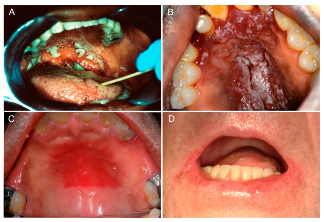
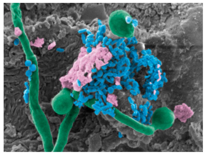
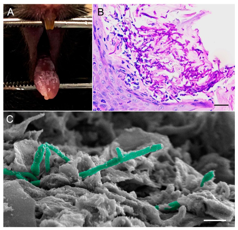
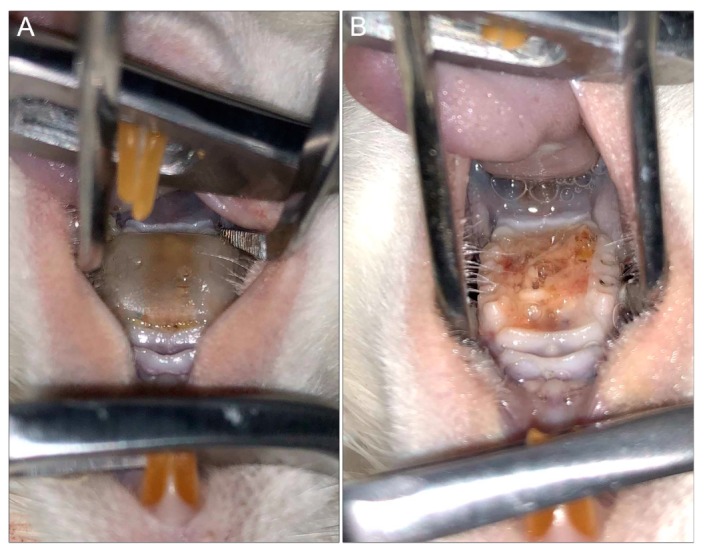
Oral candidiasis, commonly referred to as "thrush," is an opportunistic fungal infection that commonly affects the oral mucosa. The main causative agent, Candida albicans, is a highly versatile commensal organism that is well adapted to its human host; however, changes in the host microenvironment can promote the transition from one of commensalism to pathogen. This transition is heavily reliant on an impressive repertoire of virulence factors, most notably cell surface adhesins, proteolytic enzymes, morphologic switching, and the development of drug resistance. In the oral cavity, the co-adhesion of C. albicans with bacteria is crucial for its persistence, and a wide range of synergistic interactions with various oral species were described to enhance colonization in the host. As a frequent colonizer of the oral mucosa, the host immune response in the oral cavity is oriented toward a more tolerogenic state and, therefore, local innate immune defenses play a central role in maintaining Candida in its commensal state. Specifically, in addition to preventing Candida adherence to epithelial cells, saliva is enriched with anti-candidal peptides, considered to be part of the host innate immunity. The T helper 17 (Th17)-type adaptive immune response is mainly involved in mucosal host defenses, controlling initial growth of Candida and inhibiting subsequent tissue invasion. Animal models, most notably the mouse model of oropharyngeal candidiasis and the rat model of denture stomatitis, are instrumental in our understanding of Candida virulence factors and the factors leading to host susceptibility to infections. Given the continuing rise in development of resistance to the limited number of traditional antifungal agents, novel therapeutic strategies are directed toward identifying bioactive compounds that target pathogenic mechanisms to prevent C. albicans transition from harmless commensal to pathogen.

Keywords: Candida albicans; fungal–bacterial interactions; immune response; oral candidiasis.


Conflict of interest statement

Authors declare no conflict of interest.

Figures

Figure 1
Figure 1
Clinical manifestations of oral candidiasis. (A) Oropharyngeal candidiasis characterized by diffuse thick curdy white plaques that could be wiped off with gentle scraping, with extension from the soft palatal mucosa (oral candidiasis) to the oropharynx (oropharyngeal candidiasis). (B) Acute pseudomembranous candidiasis in a human immunodeficiency virus (HIV)-positive individual. Multiple coalescing raised white plaques on the hard palatal mucosa on a background of underlying diffuse erythema and hyperplasia. (C) Newton’s class II denture stomatitis clinically manifesting as diffuse erythema of the mid hard palatal mucosa in a partial denture wearer. (D) Angular cheilitis presenting as bilateral erythema and maceration of the angles of the mouth. Clinical images of consented patients attending the University of Maryland School of Dentistry.
Figure 2
Figure 2
Co-infection of Streptococcus mutans and Candida albicans. SEM pseudo-colored micrograph of a mouse tongue dorsum showing C. albicans hyphae (green) penetrating epithelial cells with S. mutans (blue) cells anchoring onto hyphae within a dense extracellular matrix (pink), 10,000x magnification.
Figure 3
Figure 3
Mouse model of oral candidiasis. (A) Diffuse white plaques with focal areas of punched-out white lesions on the tongue dorsum of a mouse depicting oral candidiasis. (B) PAS-stained image showing significant accumulation and penetration of C. albicans hyphae through the hyperkeratotic surface of the tongue dorsal epithelium. Neutrophilic Munro microabscesses in response to the infection are seen. (C) SEM micrograph, high magnification of the tongue dorsum showing C. albicans hyphae (pseudo-colored in green) penetrating epithelial cells. Bars correspond to (B) 100 µm and (C) 20 µm.
Figure 4
Figure 4
Rat model of denture stomatitis. (A) Three-dimensional (3D)-printed denture with intimate fit to the hard palatal-mucosa of a rat. (B) Denture stomatitis in a rat model demonstrating profound edema and a thick yellow biofilm of the denture-bearing hard palatal mucosa (top: tongue; bottom: hard palate).

References

    1. Millsop J.W., Faze L.N. Oral candidiasis. Clin. Dermatol. 2016;34:487–494. doi: 10.1016/j.clindermatol.2016.02.022. - DOI - PubMed
    1. Singh A., Verma R., Murari A., Agrawal A. Oral candidiasis: An overview. J. Oral Maxillofac. Pathol. 2014;18:S81–S85. - PMC - PubMed
    1. Hellstein J.W., Marek C.L. Candidiasis: Red and white manifestations in the oral cavity. Head Neck Pathol. 2019;13:25–32. doi: 10.1007/s12105-019-01004-6. - DOI - PMC - PubMed
    1. Hippocrates C.-B. Epidemics. Volume 3 Williams and Wilkins; Baltimore, MD, USA: 1939.
    1. Knoke M., Bernhardt H. The first description of an oesophageal candidosis by Bernhard von Langenbeck in 1839. Mycoses. 2006;49:283–287. doi: 10.1111/j.1439-0507.2006.01237.x. - DOI - PubMed