. 2020 Jan 16;21(2):593.
doi: 10.3390/ijms21020593.

SV40 Transfected Human Anterior Cruciate Ligament Derived Ligamentocytes-Suitable as a Human in Vitro Model for Ligament Reconstruction?

Affiliations

SV40 Transfected Human Anterior Cruciate Ligament Derived Ligamentocytes-Suitable as a Human in Vitro Model for Ligament Reconstruction?

Gundula Schulze-Tanzil et al. Int J Mol Sci. .

Abstract

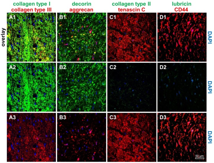
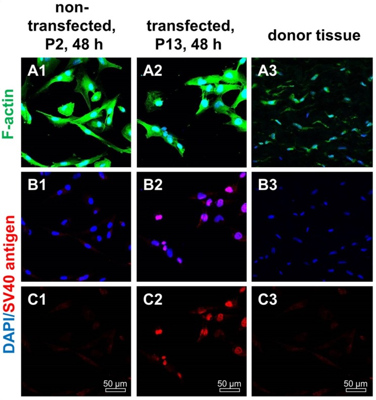
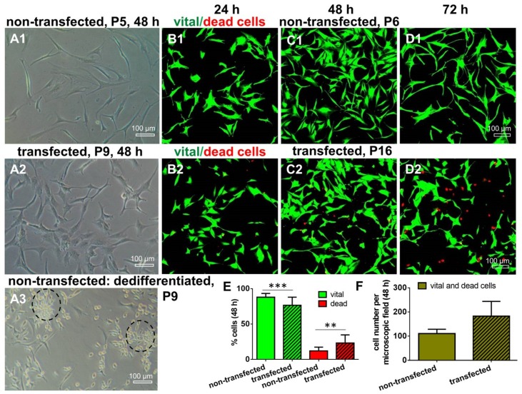
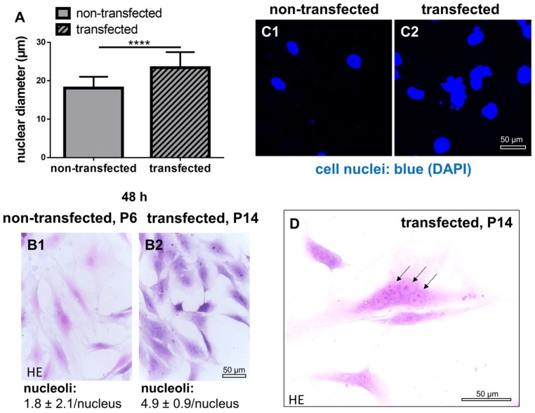
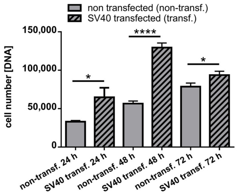
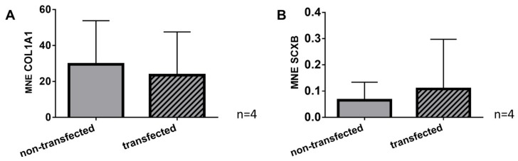
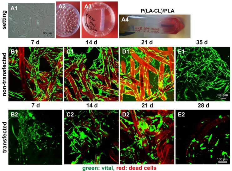
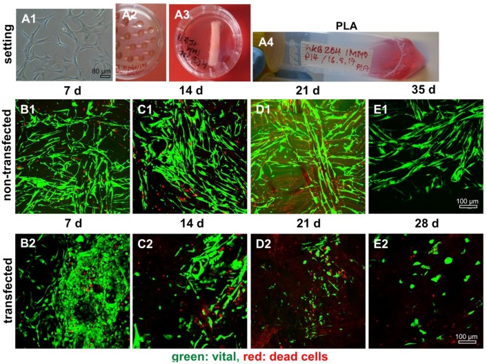
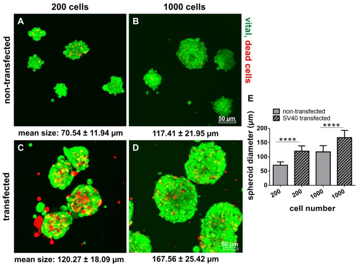
Cultured human primary cells have a limited lifespan undergoing dedifferentiation or senescence. Anterior cruciate ligaments (ACL) are hypocellular but tissue engineering (TE) requires high cell numbers. Simian virus (SV) 40 tumor (T) antigen expression could extend the lifespan of cells. This study aimed to identify cellular changes induced by SV40 expression in human ACL ligamentocytes by comparing them with non-transfected ligamentocytes and tissue of the same donor to assess their applicability as TE model. Human ACL ligamentocytes (40-year-old female donor after ACL rupture) were either transfected with a SV40 plasmid or remained non-transfected (control) before monitored for SV40 expression, survival, and DNA content. Protein expression of cultured ligamentocytes was compared with the donor tissue. Ligamentocyte spheroids were seeded on scaffolds embroidered either from polylactic acid (PLA) threads solely or combined PLA and poly (L-lactide-co-ε-caprolactone) (P(LA-CL)) threads. These scaffolds were further functionalized with fluorination and fibrillated collagen foam. Cell distribution and survival were monitored for up to five weeks. The transfected cells expressed the SV40 antigen throughout the entire observation time, but often exhibited random and incomplete cell divisions with significantly more dying cells, significantly more DNA and more numerous nucleoli than controls. The expression profile of non-transfected and SV40-positive ligamentocytes was similar. In contrast to controls, SV40-positive cells formed larger spheroids, produced less vimentin and focal adhesions and died on the scaffolds after 21 d. Functionalized scaffolds supported human ligamentocyte growth. SV40 antigen expressing ligamentocytes share many properties with their non-transfected counterparts suggesting them as a model, however, applicability for TE is limited.

Keywords: P(LA-CL); PLA; SV40; anterior cruciate ligament; embroidered scaffold; ligamentocytes; tissue engineering.


Conflict of interest statement

The authors declare no conflict of interest. The funders had no role in the design of the study; in the collection, analyses, or interpretation of data; in the writing of the manuscript, or in the decision to publish the results.

Figures

Figure 1
Figure 1
Immunohistological characterization of the human anterior cruciate ligament (ACL) donor tissue. Immunolabeling of ACL extracellular matrix components and CD44 (hyaluronan receptor) was performed using the human ACL donor tissue. (A1A3) Collagen type I (green) and type III (red), (B1B3) decorin (green) and aggrecan (red), (C1C3) collagen type II (green) and the fibroblast/ligamentocyte marker tenascin C (red), (D1D3) lubricin (green) and CD44 (red). Cell nuclei were counterstained using 4′,6′-diamidino-2-phenylindol (DAPI, blue). Scale bar: 50 µm.
Figure 2
Figure 2
F-actin and SV40 T antigen expression in non-transfected and transfected human ACL ligamentocytes after 48 h in 2D culture as well as native tissue. (A1C1) Non-transfected ligamentocytes, passage (P) 2, (A2C2) ligamentocytes transfected with the SV40 plasmid (P13) and the original donor tissue in situ (A3C3). (A1A3) Cells were stained with phalloidin-Alexa488 (green) to depict the F-actin cytoskeleton. (B1B3) Cells were immunolabeled with a specific anti-SV40 antibody (red), the cell nuclei were counterstained with 4′,6′-diamidino-2-phenylindol (DAPI, blue) (A1B3). Scale bars: 50 µm.
Figure 3
Figure 3
Survival of non-transfected and transfected human ACL ligamentocytes. (A1,A2) Images of non-transfected and transfected cells. (B1,C1,D1) Live/Dead assay of non-transfected cells and of SV40 transfected cells cultured in monolayer for 24, 48, and 72 h (B2,C2,D2). (A3) Non-transfected cells dedifferentiated in monolayer passage (P9) reflecting clusters of round proliferating cells (dashed circles). (E) Comparison of the percentage of vital (green) and dead (red) cells in non-transfected and SV40 transfected ACL ligamentocytes (48 h). (F) Comparison of the total number of cells per microscopic field after 48 h monolayer culture of non-transfected and transfected cells (n = 3, with three evaluated microscopic fields each). Scale bars: 100 µm. * p < 0.05, *** p < 0.001.
Figure 4
Figure 4
Size and shape of cell nuclei, numbers of nucleoli of non-transfected and SV40 transfected ACL ligamentocytes in 2D culture. (A) Nuclear diameter. (B1,B2) Number of nucleoli in non-transfected (B1) and transfected (B2) ACL ligamentocytes stained using hematoxylin eosin (HE). Nuclear morphology in non-transfected (C1) and transfected cells (C2). Cell nuclei of ACL ligamentocytes were visualized using 4′,6′-diamidino-2-phenylindol (DAPI, blue). (D) Random cell division without cytokinesis was shown by HE staining. Arrows: Multiple cell nuclei within one singular cell. Scale bars: 50 µm. **** p < 0.0001.
Figure 5
Figure 5
DNA content of 2D cultured non-transfected and SV40 transfected ACL ligamentocytes. Non-transfected and transfected cells were cultured for 24, 48, and 72 h and DNA content was determined by CyQuant assay. Calf thymus DNA served as a standard. Three independent experiments were performed. Transf.: transfected, * p < 0.05, **** p < 0.0001.
Figure 6
Figure 6
Expression profile of ligament ECM components and of cytoskeletal constituents in 2D cultured non-transfected and SV40 transfected ACL ligamentocytes. Collagen type I (green), actin (red) (A); decorin (green), aggrecan (red) (B); collagen type III (red) (C); tenascin C (red) (D); lubricin (green), CD44 (red) (E); fibronectin (red) (F); β1 integrin (red) (G); vinculin (green) (H); talin (red) (I); focal adhesion kinase (FAK) (red) (K); vimentin (red) (L); and α-smooth muscle actin (αSMA) (red) (M). Arrow: Extracellular deposition of tenascin C. (A1M1) Non-transfected. (A2M2) Transfected ligamentocytes. Cell nuclei were counterstained using 4′,6′-diamidino-2-phenylindol (DAPI, blue). Scale bars: 50 µm.
Figure 7
Figure 7
Gene expression profile of ligament ECM components in 2D cultured non-transfected and SV40 transfected ACL ligamentocytes. Gene expression of collagen type I (COL1A1) (A) and scleraxis (SCXB) (B). MNE: Mean normalized expression. Four independent experiments were performed.
Figure 8
Figure 8
Representative microscopic fields of non-transfected and SV40 transfected ACL ligamentocytes on P(LA-CL)/PLA scaffolds. Experimental design, cell expansion (A1); self-assembly using hanging drop method (50,000 cells per spheroid) (A2); macroscopic image of wet P(LA-CL)/PLA scaffolds (A3); and dynamical rotatory scaffold culture (A4). (B1E2) Live/Dead staining of cultures maintained up to day 35 (non-transfected cells, (B1–E1)) and day 28 (SV40 transfected cells, (B2–D2)). Vital cells: Green. Dead cell: Red. Scaffold fibers: Slightly red colored. Three independent experiments were performed. Scale bars: 100 µm.
Figure 9
Figure 9
Representative microscopic fields of non-transfected and SV40 transfected ACL ligamentocytes on PLA scaffolds. Experimental design, cell expansion (A1), self-assembly using hanging drop method (50,000 cells per spheroid) (A2), macroscopic image of the wet PLA scaffolds (A3), and dynamical rotatory scaffold culture (A4). (B1E2) Live/Dead staining of cultures up to day 35 (non-transfected) and day 28 (SV40 transfected) cells. (B1–E1) Non-transfected. (B2–E2) SV40 transfected ligamentocytes. Vital cells: Green. Dead cells: Red. Scaffold fibers: Slightly red colored. Three independent experiments were performed. Scale bars: 100 µm.
Figure 10
Figure 10
Microspheroid formation by non-transfected and SV40 transfected human ACL ligamentocytes: Spheroid size as well as cell vitality. 200 (A,C) and 1000 (B,D) human ACL ligamentocytes per spheroid either non-transfected (A,B) or SV40 transfected (C,D) were allowed to aggregate for 5 d using a spheroid multiwell plate. Live/Dead staining was performed and spheroid diameters were measured (E, 48 h). Scale bars: 50 µm. **** p < 0.0001.

References

    1. Kudo Y., Hiraoka M., Kitagawa S., Miyauchi M., Kakuo S., Zhao M., Ide T., Takata T. Establishment of human cementifying fibroma cell lines by transfection with temperature-sensitive simian virus-40 T-antigen gene and hTERT gene. Bone. 2002;30:712–717. doi: 10.1016/S8756-3282(02)00689-0. - DOI - PubMed
    1. Fujii S., Maeda H., Wada N., Kano Y., Akamine A. Establishing and characterizing human periodontal ligament fibroblasts immortalized by SV40T-antigen and hTERT gene transfer. Cell Tissue Res. 2006;324:117–125. doi: 10.1007/s00441-005-0101-4. - DOI - PubMed
    1. Fujii S., Maeda H., Wada N., Tomokiyo A., Saito M., Akamine A. Investigating a clonal human periodontal ligament progenitor/stem cell line in vitro and in vivo. J. Cell. Physiol. 2008;215:743–749. doi: 10.1002/jcp.21359. - DOI - PubMed
    1. Parkar M.H., Kuru L., O’Hare M., Newman H.N., Hughes F., Olsen I. Retroviral transduction of human periodontal cells with a temperature-sensitive SV40 large T antigen. Arch. Oral Biol. 1999;44:823–834. doi: 10.1016/S0003-9969(99)00077-1. - DOI - PubMed
    1. McNutt N.S., Culp L.A., Black P.H. Contact-inhibited revertant cell lines isolated from SV 40-transformed cells. IV. Microfilament distribution and cell shape in untransformed, transformed, and revertant Balb-c 3T3 cells. J. Cell Biol. 1973;56:412–428. doi: 10.1083/jcb.56.2.412. - DOI - PMC - PubMed

MeSH terms

LinkOut - more resources