Review
. 2020 Jan 17;9(1):253.
doi: 10.3390/jcm9010253.

Ischemia and Reperfusion Injury in Kidney Transplantation: Relevant Mechanisms in Injury and Repair

Affiliations
Review

Ischemia and Reperfusion Injury in Kidney Transplantation: Relevant Mechanisms in Injury and Repair

Gertrude J Nieuwenhuijs-Moeke et al. J Clin Med. .

Abstract

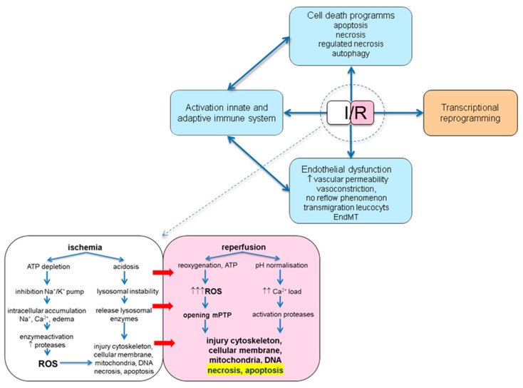
Ischemia and reperfusion injury (IRI) is a complex pathophysiological phenomenon, inevitable in kidney transplantation and one of the most important mechanisms for non- or delayed function immediately after transplantation. Long term, it is associated with acute rejection and chronic graft dysfunction due to interstitial fibrosis and tubular atrophy. Recently, more insight has been gained in the underlying molecular pathways and signalling cascades involved, which opens the door to new therapeutic opportunities aiming to reduce IRI and improve graft survival. This review systemically discusses the specific molecular pathways involved in the pathophysiology of IRI and highlights new therapeutic strategies targeting these pathways.

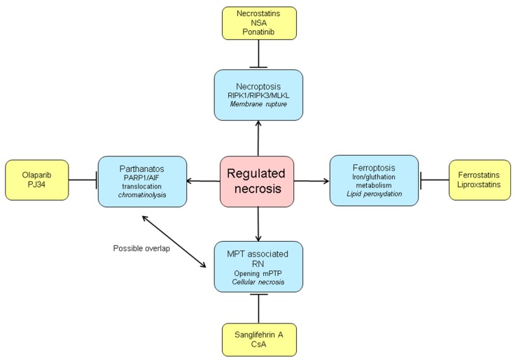
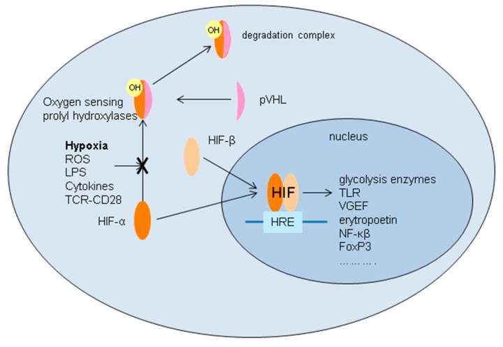
Keywords: adaptive immune system; apoptosis; delayed graft function; endothelial dysfunction; hypoxic inducible factor; innate immune system; ischemia reperfusion injury; kidney transplantation; necrosis.


Conflict of interest statement

The authors declare no conflict of interest. Parts of this review are adapted from chapter 2 of the PhD thesis: Perioperative renal protective strategies in kidney transplantation, Gertrude J. Nieuwenhuijs-Moeke, 2019.

Figures

Figure 1
Figure 1
Schematic overview of the pathophysiological consequences of ischemia and reperfusion. I/R: ischemia/reperfusion; ATP: adenosine triphosphate; EndMT: endothelial to mesenchymal transition; ROS: reactive oxygen species; mPTP: mitochondrial permeability transition pore.
Figure 2
Figure 2
Extrinsic and intrinsic apoptotic pathway. The intrinsic pathway is mediated by intracellular signals of cell stress leading to an increase in the BH3 only proteins (members of the B-cell lymphoma 2 (Bcl-2) family) resulting in an inhibition of the protectors and activation of the effectors. The effectors Bax and Bad increase the permeability of the mitochondrial membrane (MOMP: mitochondrial outer membrane permeabilisation) resulting in leakage of apoptotic proteins. One of these proteins, known as second mitochondria-derived activator of caspases (SMAC), binds to proteins that inhibit apoptosis (IAPs, by suppression of the caspase proteins) causing an inactivation of these IAPs. Another protein released from the mitochondria is cytochrome c, which binds to Apoptotic protease activating factor-1 (Apaf-1) and ATP. This complex binds to procaspase-9 creating a complex, the apoptosome. The apoptosome cleaves procaspase-9 to its active form of caspase-9, which in turn is able to activate the effector caspase-3. The extrinsic pathway is mediated through receptors of the tumor necrosis factor (TNF) receptor (TNFR) family either via the TNF path or the Fas (first apoptosis signal) path. In the TNF path binding of TNF-α to a trimeric complex of TNFR1 molecules induces activation of the intracellular death domain and the formation of the receptor-bound complex 1 made up of TNF receptor-associated death domain (TRADD), receptor-interacting protein kinase 1 (RIPK1), two ubiquitin ligases (TNFR-associated factor (TRAF)-2 and cellular inhibitors of apoptosis (clAP)1/2) and the linear ubiquitin assembly complex (LUBAC). This complex 1 can lead to a pro-survival pathway or to apoptosis. In case of apoptosis the TRADD dependant complex IIa (consisting of TRADD, Fas-associated death domain protein (FADD) and caspase-8) or the RIPK-1 dependant complex IIb also known as the ripoptosome (consisting FADD, RIPK1, RIPK3 and caspase-8) is formed. In the Fas path, presence of the Fas ligand (FasL, expressed on cytotoxic T lymphocytes) causes three Fas receptors (CD95) to trimirize. This clustering and binding to the FasL initiates binding of FADD. Three procaspase-8 or -10 molecules can then interact with the complex by their own death effector domains. The complex formed is the death-inducing signalling complex (DISC) which cleaves and activates procaspase-8 and 10. Activation of the initiator caspase-8 by both paths directly activates other members of the caspase signalling cascade such as the effector caspase-3 but also can lead to an increase in BH3-only proteins (Bim, Bid) and trigger the intrinsic pathway).
Figure 3
Figure 3
Pathways of macro-autophagy. Initiation of autophagy is regulated by mTORC1 (mammalian target of rapamycin complex 1) and AMPK (AMP-activated kinase). Together, they regulate the activity of the ULK1/2 complex consisting of ULK1/2 (Unc-51 like autophagy activating kinase), FIP200 (FAK family kinase interacting protein of 200 kDa) and the autophagy related proteins (ATG) ATG13 and ATG10. Activation of mTOR leads to the phosphorylation of this complex and inhibition of autophagy (for instance, through the phosphatidylinositol 3-kinase (PI3K)/ Protein kinase B (AKT) or the mitogen-activated protein kinase (MAPK)/ extracellular signal–regulated kinase (Erk) 1/2 signalling pathway) whereas activation of AMPK activates autophagy. AMPK, activated upon intracellular AMP increase, is able to activate autophagy by inhibition of the mTORC1 through dissociation of mTORC1 from ULK1/2 allowing ULK1/2 to be activated. AMPK, is also able to initiate autophagy in a direct way by phosphorylation of ULK1/2 forming the ULK1/2-complex.Another complex involved in the initiation is the autophagy inducible beclin-1 complex (or class III PI3K complex) which consists of Vps34 (phosphatidylinositol 3-kinase), beclin-1 (a BH3 only domain protein member of the Bcl-2 family), vps15 and ATG14. This complex is activated by the ULK-1 complex and inhibited by Bcl-2 and Bcl-XL. The ULK1/2 and class III PI3K complexes join to form the phagopore and eventually the autophagosme. This process is mediated by the ATG5-ATG12-ATG16 complex and the formation of phosphatidylethanolamine-conjugated Light Chain (LC) 3 (LC3-II) facilitating elongation of the bilipid membrane to form a closed autophagosme. The autophagosome fuses with a lysosome and the content of the autolysosome is degenerated and the components are released to be reused to synthesise new proteins or to function as an energy source for the cell. PDK-1: pyruvate dehydrogenase kinase-1.
Figure 4
Figure 4
Programs of regulated necrosis and their inhibitors. RIPK1: receptor-interacting protein kinase 1; RIPK3: receptor-interacting protein kinase 3; MLKL: Mixed Kinase Domain-Like protein; MPT: mitochondrial permeability transition; mPTP: mitochondrial permeability transition pore; RN: regulated necrosis; CsA: cyclosporin A; PARP1: poly (ADP-ribose) polymerase-1; AIF: apoptosis-inducing factor.
Figure 5
Figure 5
Interaction of leukocytes and endothelial cells in the process of transmigration of leukocytes. The increased expression of P-selectin on the endothelial cells upon I/R facilitates interaction with P-selectin glycoprotein 1 (PSGL-) expressed on the leukocytes. This results in rolling of the leukocytes on the endothelium. Subsequently, firm adherence of the leucocytes to the endothelium is achieved by interaction of lymphocyte function-associated antigen 1(LFA-1) and macrophage-1 antigen (MAC-1 or complement receptor 3, CR3) on the leukocyte and the intracellular adhesion molecule 1 (ICAM-1) on the endothelial cells. Finally, platelet endothelial cell adhesion molecule 1 (PECAM-1) facilitates transmigration of the leukocytes into the interstitial space. Once activated, these leukocytes will release several toxic substances like ROS, proteases, elastases and different cytokines in the interstitial compartment resulting in further injury like increased vascular permeability, oedema, thrombosis and parenchymal cell death.
Figure 6
Figure 6
Toll-like receptor 4 signalling. Activation of toll-like receptor 4 (TLR4) by danger associated molecular patterns (DAMPs), like high mobility group box-1 (HMGB-1), heat shock proteins (hsp) and extracellular matrix (ECM) components, leads to downstream signalling via the MyD88 (Myeloid differentiation primary-response protein 88) dependent and MyD88 independent pathway. The MyD88-dependent pathway in which MyD88 and TIRAP (toll-interleukin 1 receptor (TIR) domain containing adaptor protein) or MyD88 adapter-like (Mal) recruits and activates members of the IL-1 receptor-associated kinase (IRAK) family is considered to be the dominant pathway. IRAK activation leads to recruitment of TRAF6 (TNF receptor-associated factor 6) and subsequently activation of transforming growth factor beta-activated kinase 1 (TAK1). Activation of TAK1 then leads to the activation of inhibitor of nuclear factor-κB kinase (IKK), which results in the release of nuclear factor kappa-light-chain-enhancer of activated B cells (NF-κB) from its inhibitor, promoting translocation to the nucleus. The MyD88 independent pathway is mediated by the adapter molecules TIR-domain-containing adapter-inducing interferon-β (TRIF)/TRIF-related adaptor molecule (TRAM) and downstream signalling leads to activation of 2 inhibitor of nuclear factor-κB kinase (IKK) homologs IKKε and TANK-binding Kinase-1 (TBK1), which possibly form a complex together and activate transcription factors NF-κB and IFN-regulatory factor 3 (IRF3). From here, proinflammatory gene transcription is initiated. TLR4 signalling is inhibited by Eritoran and TJ-M2010-2.
Figure 7
Figure 7
Routes of the complement system with its inhibitors currently studied in kidney transplantation. Damps released upon I/R are able to activate all three pathways via binding to C1q (classical pathway), C3 (alternative pathway) or pattern recognition receptors (PRRs) of the lectin path. All activating routes converge and lead to the formation of the complement component (C) 3 (C3) convertase (C4b2b, C3bBbP). C3 convertase cleaves and activates additional C3, creating C3a and C3b. C3b together with C4b2b forms the C5 convertase, which will cleave C5 into C5a and C5b. C5b together with C6–9 will then form the Membrane Attack Complex (MAC, C5b-9). The formed complement effectors will lead to opsonisation (C3b), chemotaxis of neutrophils and macrophages (C3a, C5a). The formed MAC inserted into the cellular membrane is associated with a proinflammatory response via noncanonical NF-ΚB signalling. C1-inhibitors (C1-INH), Cinryze® and Berinert® target complement initiation and APT070 complement amplification. Eculizumab and Tesidolumab inhibit complement activation at the level of C5.
Figure 8
Figure 8
Interaction of the innate and adaptive immune system in the pathophysiology of ischemia and reperfusion injury. DAMPs released upon I/R are able to activate the innate immune system by binding to PRRs like complement receptors and TLRs. Activation of these receptors will lead to production of pro-inflammatory cytokines and chemokines and chemotaxis, opsonisation and activation of leucocytes like macrophages, neutrophils and natural killer (NK) cells. Additionally, immature dendritic cells can be activated, which, after maturation, are able to activate the adaptive immune system in a direct manner by antigen presentation to B- and T-cells or indirectly via cytokine signalling. Treg: regulatory T-cell.
Figure 9
Figure 9
Intracellular stabilisation and activation of hypoxic inducible factor. Under normoxemic conditions, proline on the hypoxic inducible factor (HIF) α (HIFα) subunit is rapidly hydroxylated by oxygen-sensing prolyl hydroxylase (PHD). This induces a conformational change enabling von Hippel–Lindau tumour suppressor protein (pVHL) to bind with the α-subunit, leading to degradation of the HIF-α subunit. Ischemia (or other signals like lipopolysaccharide (LPS), various cytokines, etc.) will lead to inhibition of the oxygen-dependent PHD, enabling nuclear translocation of the α subunit, binding of the α and β subunit and formation of HIF. In the nucleus, HIF binds with the hypoxia response promotor element (HRE) leading to the transcription of various genes. VGEF: vascular endothelial growth factor.

Similar articles

Cited by

References

    1. World Health Organisation . Disease Burden and Mortality Estimates. WTO; Geneva, Switzerland: 2015. [(accessed on 15 January 2020)]. Available online: https://www.who.int/healthinfo/global_burden_disease/estimates/en/index1....
    1. Liyanage T., Ninomiya T., Jha V., Neal B., Patrice H.M., Okpechi I., Zhao M.H., Lv J., Garg A.X., Knight J., et al. Worldwide access to treatment for end-stage kidney disease: A systematic review. Lancet. 2015;385:1975–1982. doi: 10.1016/S0140-6736(14)61601-9. - DOI - PubMed
    1. Global Observatory on Donation and Transplantation. [(accessed on 15 January 2020)];2019 Available online: http://www.transplant-observatory.org.
    1. Cooper J.T., Chin L.T., Krieger N.R., Fernandez L.A., Foley D.P., Becker Y.T., Odorico J.S., Knechtle S.J., Kalayoglu M., Sollinger H.W., et al. Donation after cardiac death: The university of Wisconsin experience with renal transplantation. Am. J. Transplant. 2004;4:1490–1494. doi: 10.1111/j.1600-6143.2004.00531.x. - DOI - PubMed
    1. Koffman G., Gambaro G. Renal transplantation from non-heart-beating donors: A review of the European experience. J. Nephrol. 2003;16:334–341. - PubMed

LinkOut - more resources