. 2020 Jan 17;12(1):233.
doi: 10.3390/cancers12010233.

Elevated Expression of Lumican in Lung Cancer Cells Promotes Bone Metastasis through an Autocrine Regulatory Mechanism

Affiliations

Elevated Expression of Lumican in Lung Cancer Cells Promotes Bone Metastasis through an Autocrine Regulatory Mechanism

Kuan-Chung Hsiao et al. Cancers (Basel). .

Abstract

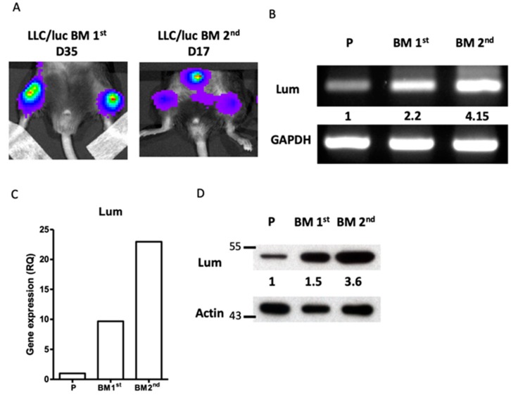
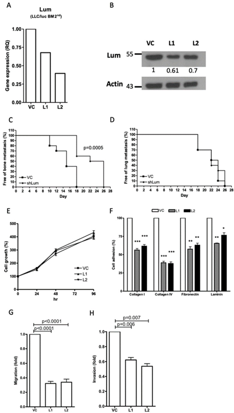
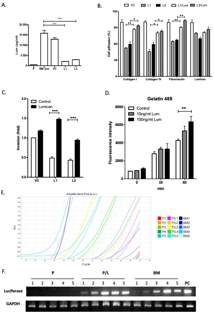
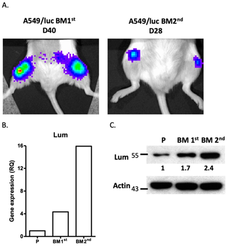
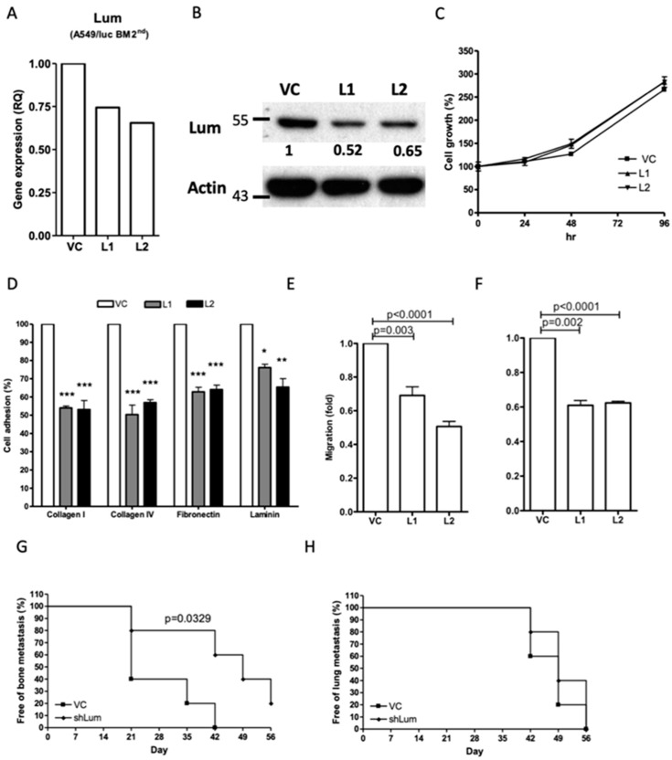
Background: The microarray analysis of whole-genome expression indicated that the gene encoding the protein lumican, which is associated with extracellular matrix (ECM) interaction, was highly expressed in osteotropic lung cancer cell lines with an enhanced capacity of bone metastasis. Methods: The expression of lumican in the osteotropic lung cancer cells was downregulated, and the in vitro migration, invasion, and adhesion of cancer cells to ECM components, and the in vivo bone metastasis capacity of these cells were examined. Exogenous lumican was provided to study the autocrine regulation mechanism of lumican in the bone metastasis of lung cancer cells. Results: Transfection with lumican-specific short hairpin RNA (shRNA) in the osteotropic lung cancer cells reduced the establishment of in vivo bone metastasis, but not lung metastasis. Reduction in the expression of lumican also decreased the attachment of lung osteotropic cancer cells to several components of the ECM and suppressed cell migration and invasion in vitro. Exogenous lumican restored these reduced capacities of lumican knockdown cells and promoted the seeding of lung cancer cells in the bone microenvironment. Conclusions: These results suggested that lumican promotes the metastasis of lung cancer cells to the bones via an autocrine regulatory mechanism, and blocking this interaction may provide a new therapeutic approach to reduce bone metastasis in cases of lung cancer.

Keywords: bone metastasis; lumican; lung cancer.


Conflict of interest statement

The authors declare no conflict of interest.

Figures

Figure 1
Figure 1
Expression of lumican in osteotropic LLC/luc cells. (A) To develop lung cancer cells with a higher capacity of bone metastasis, a mouse LLC lung cancer cell line transfected with the luciferase gene (LLC/luc) was injected into the left ventricle of a C57BL/6 mouse. After 35 days (D35), the luciferase activity was observed in the femurs of mice by the in vivo imaging system(IVIS). The bone marrow cells of mice with bone metastases were collected and cultured in vitro to establish the first bone metastatic cell line, LLC/luc BM 1st. The BM 1st cells were injected again into a different mouse and the luciferase activity was detected on D17. The bone marrow cells of this mouse were collected and cultured in vitro to establish a second cell line exhibiting high bone metastasis, LLC/luc BM 2nd. The expression of lumican in the parental LLC/luc (P), LLC/luc BM 1st, and LLC/luc BM 2nd cells was determined by RT-PCR (B), quantitative-real-time PCR (C), and Western blot analysis (D). The level of lumican expression in each cell was individually normalized to the internal control (GAPDH or actin), and the numbers in (B,D) indicate the expression levels of lumican in the bone metastatic LLC/luc cells as compared to those in the parental LLC/luc cells (level set to 1).
Figure 2
Figure 2
Effect of lumican knockdown on the function of bone metastatic LLC/luc BM 2nd cells. The expression of lumican in LLC/luc BM 2nd cells transfected with a control vector (VC) and a lumican-specific short hairpin RNA (shRNA) plasmid (L1 and L2) was determined by real-time RT-PCR (A) and Western blot analysis (B). The level of lumican expression in each cell was individually normalized to the internal control (actin), and the numbers in (B) indicate the level of lumican expression in lumican knockdown LLC/luc BM 2nd cells as compared to that in the cells transfected with the control vector. The LLC/luc BM 2nd cells transfected with a control vector (VC) and a lumican-specific shRNA (shLum) were administered by injecting them intracardiac (I.C.) and intravenous (I.V.) to allow the establishment of bone (C) and lung (D) metastasis (n = 10, from two separate experiments), respectively. The presence of tumor metastasis as determined by the presence of luciferase activity was detected by the IVIS imaging system. The cell proliferation (E) and adhesion to the extracellular matrix (ECM) components (F) of LLC/luc BM 2nd cells transfected with the control vector (VC) and the lumican-specific shRNA plasmid (L1, L2) were determined by an Methylthiazolyldiphenyl-tetrazolium bromide (MTT) assay and a cell adhesion assay, respectively. The migration (G) and invasion (H) abilities of LLC/luc BM 2nd cells transfected with the control vector and the lumican-specific shRNA plasmid were determined by the Transwell migration assay. * p ≤ 0.05, ** p ≤ 0.01, and *** p ≤ 0.001. The error bars are defined as means ± SEM.
Figure 3
Figure 3
Effect of exogenous lumican on function of lumican knockdown bone metastatic LLC/luc cells. (A) The level of lumican in the culture supernatant of the parental LLC/luc (P), LLC/luc BM 2nd (BM 2nd), and LLC/luc BM 2nd cells transfected with a control and a lumican-specific shRNA vector was determined by an ELISA assay. (B) Cell adhesion to different ECM components was determined in the bone metastatic cells transfected with a control vector, LLC/luc cells (VC), and lumican knockdown LLC/luc BM 2nd cells incubated with (L1/lum and L2/lum) and without (L1 and L2) the recombinant lumican protein. (C) Cell invasion was determined in LLC/luc BM 2nd cells transfected with a control (VC) and a lumican-specific shRNA vector (L1 and L2) after incubation with (lumican) and without (control) the recombinant lumican protein, respectively. (D) Lumican knockdown LLC/luc BM 2nd cells were cultured in a medium containing the recombinant lumican protein (10 ng/mL and 100 ng/mL) at 37 °C. In total, 100 μL of the culture supernatant was collected after intervals of 30 min and incubated with fluorescein-conjugated gelatin as a substrate for matrix metalloproteinase 2/9 (MMP2/9). The fluorescence released from the degraded substrates was then measured. (E) The LLC/luc parental cells co-cultured with (P/L1 to P/L5) and without (P1 to P5) recombinant lumican protein and LLC/luc BM 2nd cells (BM1 to BM5) were injected into mice (n = 5) I.C. The total RNA of bone marrow cells was extracted one day after injecting them I.C. The expression of luciferase gene was determined by quantitative-real-time PCR to detect the presence of tumor cells in the bone marrow, and the amplification of luciferase gene was confirmed by agarose gel electrophoresis (F). GAPDH was used as an internal control. The DNA extracted from the parental LLC/luc cells was used as a positive control (PC). * p ≤ 0.05, ** p ≤ 0.01, and *** p ≤ 0.001.
Figure 4
Figure 4
Expression of lumican in bone metastatic A549/luc cells. (A) The A549 human lung cancer cell line transfected with the luciferase gene (A549/luc) was injected into the left ventricle of a NOD-SCID mouse. After 40 days (D40), the luciferase activity was observed in the femurs of mice by the IVIS imaging system. The bone marrow cells of mice with bone metastasis were collected and cultured in vitro to establish the bone metastatic cell line, A549/luc BM 1st. The BM 1st cells were injected again into a different mouse and the luciferase activity was detected on D28. The bone marrow cells of this mouse were collected and cultured in vitro to establish a highly bone metastatic cell line, A549/luc BM 2nd. The expression of lumican in the parental A549/luc (P), A549/luc BM 1st, and A549/luc BM 2nd cells was determined by quantitative-real-time PCR (B) and Western blot analysis (C). The level of lumican expression in each cell line was individually normalized to the internal control (actin), and the numbers in (C) indicate the level of lumican expression in bone metastatic A549/luc cells as compared to the parental A549/luc cells (level set to 1).
Figure 5
Figure 5
Effect of lumican knockdown on cellular function of bone metastatic A549/luc cells. The expression of lumican in bone metastatic A549/luc BM 2nd cells transfected with a control (VC) and a lumican-specific shRNA vector (L1 and L2) was determined by real-time RT-PCR (A) and Western blot analysis (B). The level of lumican expression in each cell was individually normalized to the internal control (actin), and the numbers indicate the level of lumican expression in lumican knockdown A549/luc BM 2nd cells as compared to that in the cells transfected with the control vector. The cell growth (C) and ECM component adhesion (D) of A549/luc BM 2nd cells transfected with a control and a lumican-specific shRNA vector was determined by an MTT assay and a cell adhesion assay. The migration (E) and invasion (F) abilities of A549/luc BM 2nd cells transfected with the control and the lumican-specific shRNA vector were determined by the Transwell migration assay. The A549/luc BM 2nd cells transfected with a control (VC) and a lumican-specific shRNA (shLum) vector were administered by intracardiac and intravenous injection to allow the establishment of the bone (G) and lung (H) metastasis (n = 5), respectively. The presence of tumor metastasis, as determined by the presence of luciferase activity, was detected by the IVIS imaging system. * p ≤ 0.05, ** p ≤ 0.01, and *** p ≤ 0.001. The error bars are defined as means ± SEM.

References

    1. Spano D., Heck C., De Antonellis P., Christofori G., Zollo M. Molecular networks that regulate cancer metastasis. Semin. Cancer Biol. 2012;22:234–249. doi: 10.1016/j.semcancer.2012.03.006. - DOI - PubMed
    1. Herbst R.S., Heymach J.V., Lippman S.M. Lung cancer. N. Engl. J. Med. 2008;359:1367–1380. doi: 10.1056/NEJMra0802714. - DOI - PMC - PubMed
    1. Langer C.J. Role of zoledronic acid in the setting of bone metastases from non-small-cell lung cancer. Clin. Lung Cancer. 2005;6:314–316. doi: 10.3816/CLC.2005.n.012. - DOI - PubMed
    1. Tsuya A., Kurata T., Tamura K., Fukuoka M. Skeletal metastases in non-small cell lung cancer: A retrospective study. Lung Cancer. 2007;57:229–232. doi: 10.1016/j.lungcan.2007.03.013. - DOI - PubMed
    1. Al Husaini H., Wheatley-Price P., Clemons M., Shepherd F.A. Prevention and management of bone metastases in lung cancer: A review. J. Thorac. Oncol. Off. Publ. Int. Assoc. Study Lung Cancer. 2009;4:251–259. doi: 10.1097/JTO.0b013e31819518fc. - DOI - PubMed