. 2020 Jan 17;9(1):33.
doi: 10.3390/antibiotics9010033.

Inhibitory Activity of a Scorpion Defensin BmKDfsin3 against Hepatitis C Virus

Affiliations

Inhibitory Activity of a Scorpion Defensin BmKDfsin3 against Hepatitis C Virus

Yuting Cheng et al. Antibiotics (Basel). .

Abstract

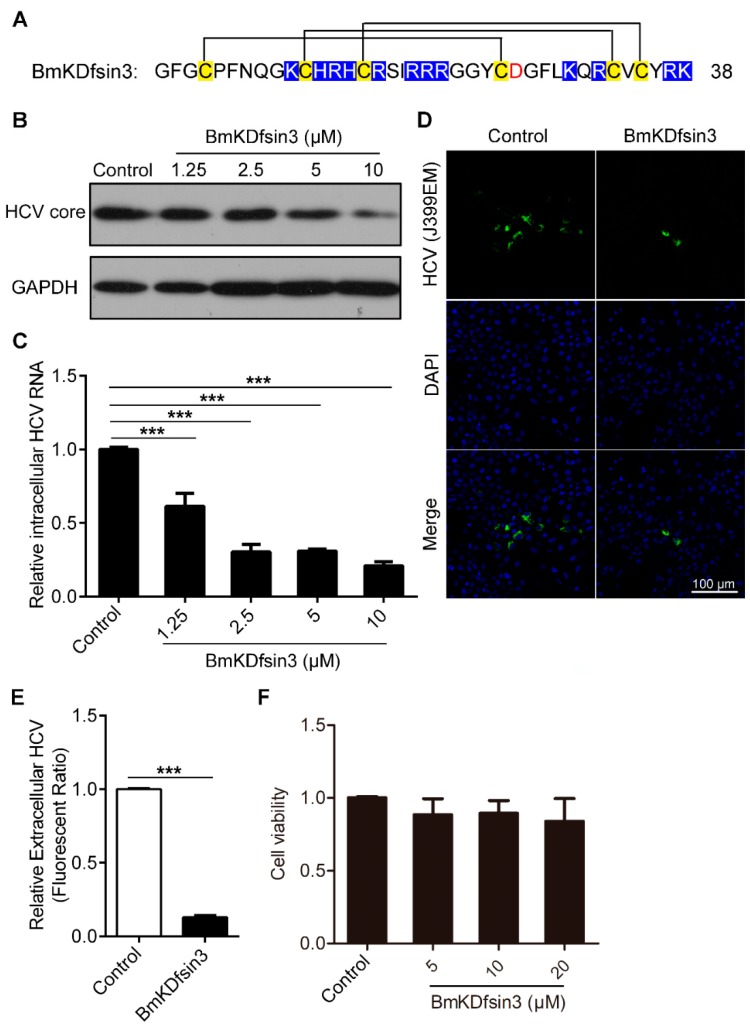
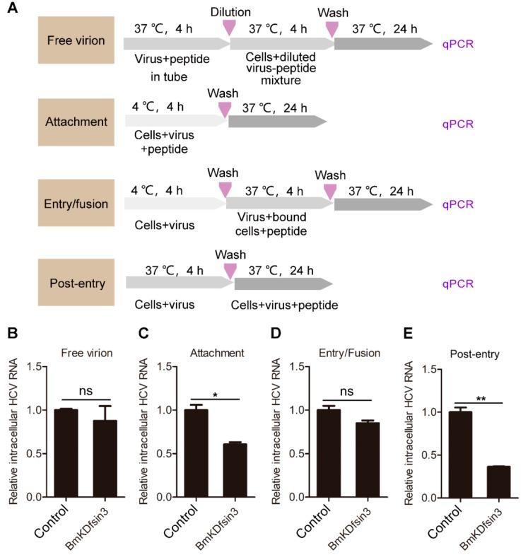
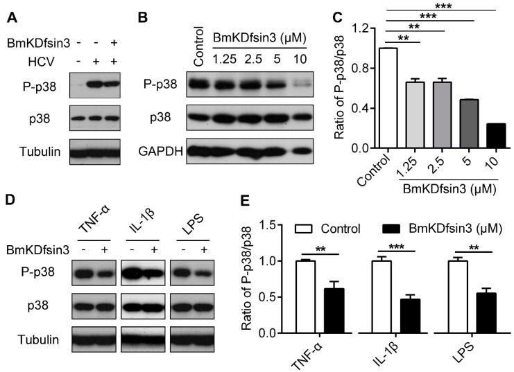
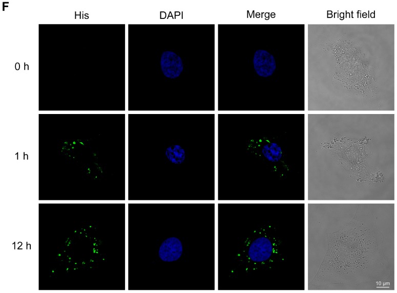
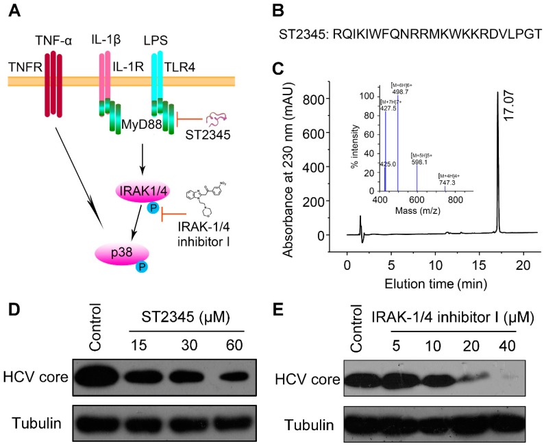
Hepatitis C virus (HCV) infection is a major worldwide health problem which can cause chronic hepatitis, liver fibrosis and hepatocellular carcinoma (HCC). There is still no vaccine to prevent HCV infection. Currently, the clinical treatment of HCV infection mainly relies on the use of direct-acting antivirals (DAAs) which are expensive and have side effects. Here, BmKDfsin3, a scorpion defensin from the venom of Mesobuthus martensii Karsch, is found to dose-dependently inhibit HCV infection at noncytotoxic concentrations and affect viral attachment and post-entry in HCV life cycle. Further experimental results show that BmKDfsin3 not only suppresses p38 mitogen-activated protein kinase (MAPK) activation of HCV-infected Huh7.5.1 cells, but also inhibits p38 activation of Huh7.5.1 cells stimulated by tumor necrosis factor-α (TNF-α), interleukin-1β (IL-1β) or lipopolysaccharide (LPS). BmKDfsin3 is also revealed to enter into cells. Using an upstream MyD88 dimerization inhibitor ST2345 or kinase IRAK-1/4 inhibitor I, the inhibition of p38 activation represses HCV replication in vitro. Taken together, a scorpion defensin BmKDfsin3 inhibits HCV replication, related to regulated p38 MAPK activation.

Keywords: Hepatitis C virus (HCV); p38; scorpion defensin.


Conflict of interest statement

The authors declare no competing financial interest.

Figures

Figure 1
Figure 1
BmKDfsin3 inhibits HCV replication in vitro under noncytotoxic concentrations. (A) Amino acid sequence of BmKDfsin3. There are six cysteine residues forming three pairs of disulfide bonds. Cysteine residues are labeled with yellow, basic residues with blue, and acidic residues with red. (B,C) Concentration-dependent inhibition of BmKDfsin3 on HCV infection in Huh7.5.1 cells. Huh7.5.1 cells were preincubated with the BmKDfsin3 at different concentrations for 1 h and then infected with HCV J399EM at an multiplicity of infection (MOI) of 0.1. After 72 h, cells were collected, and intracellular HCV core protein levels were analyzed by western blotting (B) and intracellular HCV RNA was analyzed by qPCR (C). The J399EM was derived from the JFH-1 virus (HCV genotype 2a strain) by insertion of eGFP into the HCV NS5A region. (D) Inhibition of BmKDfsin3 on extracellular virus particles of Huh7.5.1 cells. Huh 7.5.1 cells treated with or without BmKDfsin3 (5 μM) were infected by J399EM for 72 h, and then supernatant was collected and used to incubate naïve Huh7.5.1 cells for 72 h. Virus in Huh 7.5.1 cells were observed by immunofluorescence microscope. HCV, green. 4’,6-diamidino-2-phenylindole (DAPI), blue. Scale bar, 100 μm. (E) The statistics of the fluorescence ratio of extracellular virus particles as described in (D). (F) Cytotoxicity of BmKDfsin3 to Huh7.5.1 cells by the MTT assay. BmKDfsin3 was dissolved in the medium and the medium without BmKDfsin3 was used as a negative control in all experiments. The internal control of subfigure (C) was glyceraldehyde-phosphate dehydrogenase (GAPDH). ***, p < 0.001. Data represented the mean ± standard deviation (SD) of at least three independent experiments.
Figure 2
Figure 2
BmKDfsin3 affects the attachment and post-entry stages during the HCV infection cycle. (A) Schematic diagram for studying the action stage of BmKDfsin3 on HCV. (B–E) The effects of BmKDfin3 on free virion (B), attachment (C), entry/fusion (D), and post-entry (E) stages of HCV in Huh7.5.1 cells. Huh7.5.1 cells were infected with J399EM at an MOI of 0.1 and treated with BmKDfsin3 as described in (A). All experiments were detected by qPCR. BmKDfsin3 was dissolved in the medium and the medium without BmKDfsin3 was used as a negative control. The internal controls of subfigures (BE) were GAPDH. ns, no significance. *, p < 0.05. **, p < 0.01. Data represented the mean ± SD of at least three independent experiments.
Figure 3
Figure 3
BmKDfsin3 inhibits p38 activation. (AC) BmKDfsin3 suppresses p38 phosphorylation in HCV-infected Huh7.5.1 cells. Huh7.5.1 cells were infected with J399EM at an MOI of 1 for 2 h, and then treated with BmKDfsin3 (5 μM) following the detection of intracellular total p38, phosphorylated p38 and HCV core protein levels by western blotting (A). Huh7.5.1 cells were treated as (A) and then with the BmKDfsin3 at different concentrations. The expression levels of total p38 and phosphorylated p38 were determined by western blotting (B). Grayscale of Figure B was analyzed with Image J software (C). BmKDfsin3 was dissolved in the medium and the medium without BmKDfsin3 was used as a negative control. (D,E) Effect of BmKDfsin3 on p38 activation in Huh7.5.1 cells stimulated by TNF-α, IL-1β or LPS. Huh7.5.1 cells were incubated with TNF-α (100 ng/mL), IL-1β (1 ng/mL), and LPS (1 μg/mL) (E) for 2 h, and then treated by BmKDfsin3 (5 μM). Phosphorylation level of p38 was detected by western blotting. Grayscale of Figure D was analyzed with Image J software (E). TNF-α, IL-1β, and LPS were dissolved in phosphate buffered saline (PBS) and PBS was used as a negative control. **, p < 0.01. ***, p < 0.001. Data represented the mean ± SD of at least three independent experiments. (F) The entry of His-BmKDfsin3 to Huh7.5.1 cells. Cells were treated with His-BmKDfsin3 (5 μM) for 0 h, 1 h and 12 h, respectively, and then stained with anti-His antibody and DAPI. Cells were observed using a confocal microscopy. His-BmKDfsin3, green. DAPI, blue. Scale bar, 10 μm. Cells were treated with His-BmKDfsin3 (5 μM) for 0 h as a control. The subfigure (F) was representative of at least ten independent pictures.
Figure 3
Figure 3
BmKDfsin3 inhibits p38 activation. (AC) BmKDfsin3 suppresses p38 phosphorylation in HCV-infected Huh7.5.1 cells. Huh7.5.1 cells were infected with J399EM at an MOI of 1 for 2 h, and then treated with BmKDfsin3 (5 μM) following the detection of intracellular total p38, phosphorylated p38 and HCV core protein levels by western blotting (A). Huh7.5.1 cells were treated as (A) and then with the BmKDfsin3 at different concentrations. The expression levels of total p38 and phosphorylated p38 were determined by western blotting (B). Grayscale of Figure B was analyzed with Image J software (C). BmKDfsin3 was dissolved in the medium and the medium without BmKDfsin3 was used as a negative control. (D,E) Effect of BmKDfsin3 on p38 activation in Huh7.5.1 cells stimulated by TNF-α, IL-1β or LPS. Huh7.5.1 cells were incubated with TNF-α (100 ng/mL), IL-1β (1 ng/mL), and LPS (1 μg/mL) (E) for 2 h, and then treated by BmKDfsin3 (5 μM). Phosphorylation level of p38 was detected by western blotting. Grayscale of Figure D was analyzed with Image J software (E). TNF-α, IL-1β, and LPS were dissolved in phosphate buffered saline (PBS) and PBS was used as a negative control. **, p < 0.01. ***, p < 0.001. Data represented the mean ± SD of at least three independent experiments. (F) The entry of His-BmKDfsin3 to Huh7.5.1 cells. Cells were treated with His-BmKDfsin3 (5 μM) for 0 h, 1 h and 12 h, respectively, and then stained with anti-His antibody and DAPI. Cells were observed using a confocal microscopy. His-BmKDfsin3, green. DAPI, blue. Scale bar, 10 μm. Cells were treated with His-BmKDfsin3 (5 μM) for 0 h as a control. The subfigure (F) was representative of at least ten independent pictures.
Figure 4
Figure 4
Inhibition of p38 activation suppresses HCV replication in vitro. (A) Schematic diagram of the p38 MAPK signal pathway and its inhibiting by ST2345 (MyD88 inhibitor) and IRAK-1/4 inhibitor I (IRAK1 and IRAK4 inhibitor). (B) Amino acid sequence of ST2345. (C) Mass spectrometry and HPLC analysis of ST2345 (1 μg/μL). (D,E) The inhibition of ST2345 and IRAK-1/4 inhibitor I on HCV infection. Huh7.5.1 cells were infected with J399EM at an MOI of 0.1, and then treated with different concentrations of ST2345 (D) and IRAK-1/4 inhibitor I (E) for 72 h, respectively. The expression level of the HCV core protein was determined by western blotting. The ST2345 peptide was dissolved in medium and the medium without ST2345 was used as a negative control. The IRAK-1/4 inhibitor I was dissolved in dimethyl sulfoxide (DMSO) and the DMSO without IRAK-1/4 inhibitor I was used as a negative control.

Similar articles

Cited by

References

    1. Mansour S.C., Pena O.M., Hancock R.E. Host defense peptides: Front-line immunomodulators. Trends Immunol. 2014;35:443–450. doi: 10.1016/j.it.2014.07.004. - DOI - PubMed
    1. Holly M.K., Diaz K., Smith J.G. Defensins in viral infection and pathogenesis. Annu. Rev. Virol. 2017;4:369–391. doi: 10.1146/annurev-virology-101416-041734. - DOI - PubMed
    1. Smith J.G., Nemerow G.R. Mechanism of adenovirus neutralization by human alpha-defensins. Cell Host Microbe. 2008;3:11–19. doi: 10.1016/j.chom.2007.12.001. - DOI - PubMed
    1. Lei R., Hou J., Chen Q., Yuan W., Cheng B., Sun Y., Jin Y., Ge L., Ben-Sasson S.A., Chen J., et al. Self-assembling myristoylated human alpha-defensin 5 as a next-generation nanobiotics potentiates therapeutic efficacy in bacterial infection. ACS Nano. 2018;12:5284–5296. doi: 10.1021/acsnano.7b09109. - DOI - PubMed
    1. Xu D., Liao C., Zhang B., Tolbert W.D., He W., Dai Z., Zhang W., Yuan W., Pazgier M., Liu J., et al. Human enteric alpha-defensin 5 promotes shigella infection by enhancing bacterial adhesion and invasion. Immunity. 2018;48:1233–1244. doi: 10.1016/j.immuni.2018.04.014. - DOI - PMC - PubMed

LinkOut - more resources