. 2020 Jan 17;13(2):452.
doi: 10.3390/ma13020452.

Cytotoxicity of Self-Etch Versus Etch-and-Rinse Dentin Adhesives: A Screening Study

Affiliations

Cytotoxicity of Self-Etch Versus Etch-and-Rinse Dentin Adhesives: A Screening Study

Luisa Fröb et al. Materials (Basel). .

Abstract

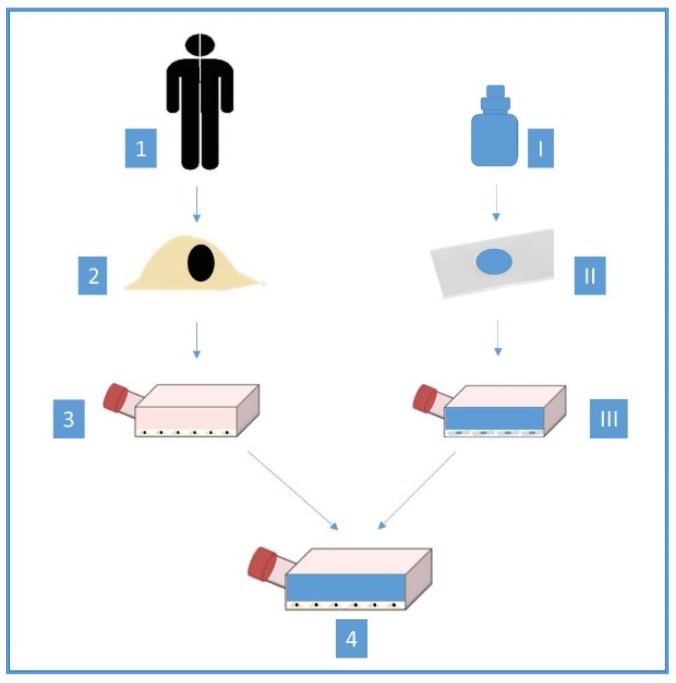
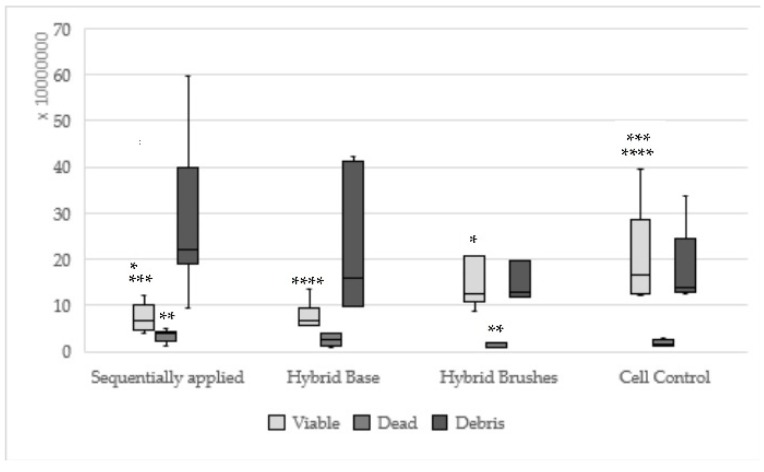
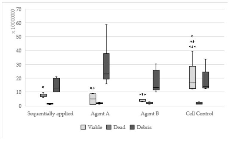
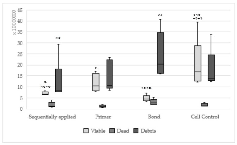
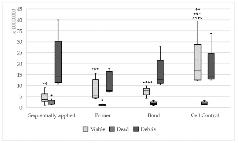
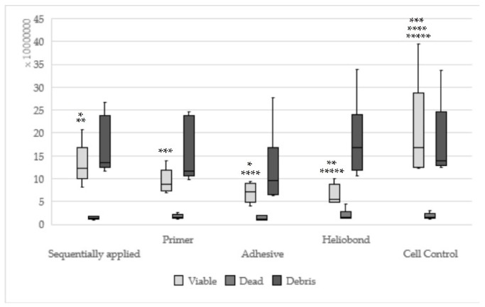
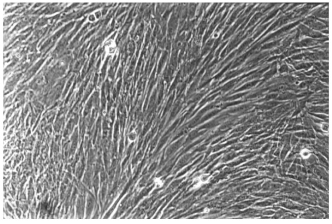
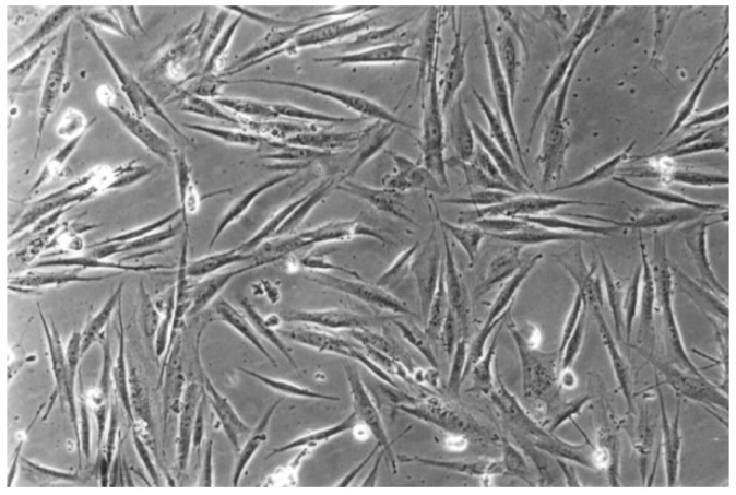
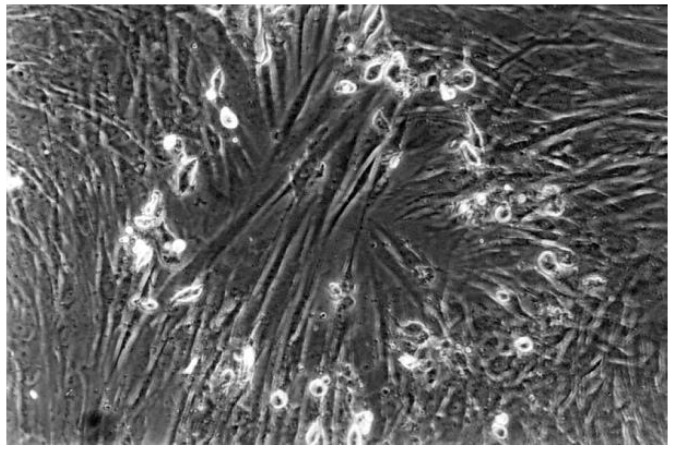
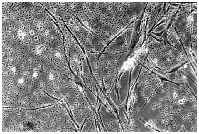
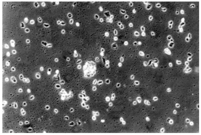
Six dentin adhesives were tested in vitro regarding their cytotoxicity on human fibroblasts. The adhesives Hybrid Bond, One-up Bond F Plus, AdheSE, Clearfil SE Bond, Optibond Solo Plus and Syntac were eluted with culture medium as single or sequentially applied adhesive part for 24 h. 75 Petri dishes were produced per group. They were evaluated triangulated, comprising the quantitative evaluation (105 ones) to determine "viable", "dead" and "debris" cells with the use of a cell-counter and the reactivity index was also identified based on the qualitative assessment (420 ones). One-up Bond F Plus, AdheSE and Clearfil SE Bond showed a statistical difference of viable cells to the cell control. For One-up Bond F Plus, statistically, differences compared to hybrid bond and Syntac were also found. All the adhesives except One-up Bond F Plus showed significant differences between single and sequentially applied adhesive part regarding the quantitative evaluation. The test material showed a moderate grade of cytotoxicity. As a result, a statistically significant difference of the cytotoxicity between the self-etch and etch-and-rinse adhesives cannot be demonstrated regarding the qualitative evaluation and the reactivity index, but the differences between sequentially applied and single applied components can be proved.

Keywords: cells; cytotoxicity; dentin adhesives; fibroblasts; in vitro; screening.


Conflict of interest statement

The authors declare no conflict of interest.

Figures

Figure 1
Figure 1
The method of the cytotoxicity assay (1 = biopsy of normal human gingival tissue, 2 = isolation of fibroblast cells, 3 = cultivation of fibroblast cells, I = different dentin adhesives, II = adhesives were applied centrally to the sterile bases of glass slides, III = eluate was obtained, 4 = eluate added to fibroblasts in Petri dishes).
Figure 2
Figure 2
Results of hybrid bond sequentially applied, single applied (Hybrid base, hybrid brushes) and cell control; (sequentially applied vs. hybrid brushes * viable: p = 0.008 ** dead: p = 0.003; same asterisks show significant differences in the corresponding groups).
Figure 3
Figure 3
Results of One-up Bond F Plus sequentially applied, single applied (Agent A, Agent B) and Cell control (same asterisks show significant differnces in the corresponding groups).
Figure 4
Figure 4
Results of AdheSE sequentially applied, single applied (Primer, Bond) and Cell control; (Sequentially applied vs. AdheSE Primer * viable: p = 0.008; sequentially applied vs. AdheSE Bond ** debris: p = 0.03; same asterisks show significant differnces in the corresponding groups).
Figure 5
Figure 5
Results of Clearfil SE Bond sequentially applied, single applied (Primer, Bond) and Cell control; (Sequentially applied vs. Clearfil SE Bond Primer * dead: p = 0.04; same asterisks show significant differnces in the corresponding groups).
Figure 6
Figure 6
Results of Syntac sequentially applied, single applied (Primer, Adhesives, Heliobond) and Cell control; (Sequentially applied vs. Adhesive * viable: p = 0.005, sequentially applied vs. Heliobond ** viable: p = 0.002; same asterisks show significant differnces in the corresponding groups).
Figure 7
Figure 7
The cell control (No. 7) presents regular dense of fibroblasts with characteristic long cells and normal mitoses (100-fold magnification).
Figure 8
Figure 8
With Clearfil SE Bond (No. 4), the cell culture appears less dense than the cell control. Normal cells are predominating, but few rounded cells are found (100-fold magnification).
Figure 9
Figure 9
The contact cells to the material One-up Bond F Plus (No. 2) are rounded or dead. The fibroblast lawn is less dense than control cultures (100-fold magnification).
Figure 10
Figure 10
The cell culture exposed to dentin Adhesive Syntac (No. 5) is much less dense than the cell control, with many rounded and dead cells (100-fold magnification).
Figure 11
Figure 11
All fibroblasts under the influence of Optibond Solo Plus (No. 6) are in the process of dying. Many rounded and dead fibroblasts found already in the cell culture (100-fold magnification).

Similar articles

Cited by

References

    1. Van Meerbeek B., Yoshihara K., Yoshida Y., Mine A., de Munck J., van Landuyt K.L. State of the art of self-etch adhesives. Dent. Mater. 2011;27:17–28. doi: 10.1016/j.dental.2010.10.023. - DOI - PubMed
    1. Giannini M., Makishi P., Ayres A.P.A., Vermelho P.M., Fronza B.M., Nikaido T., Tagami J. Self-Etch Adhesive Systems: A Literature Review. Braz. Dent. J. 2015;26:3–10. doi: 10.1590/0103-6440201302442. - DOI - PubMed
    1. Szep S., Kunkel A., Ronge K., Heidemann D. Cytotoxicity of modern dentin adhesives--in vitro testing on gingival fibroblasts. J. Biomed. Mater. Res. 2002;63:53–60. doi: 10.1002/jbm.10083. - DOI - PubMed
    1. Sangwichit K., Kingkaew R., Pongprueksa P., Senawongse P. Effect of thermocycling on the durability of etch-and-rinse and self-etch adhesives on dentin. Dent. Mater. J. 2016;35:360–368. doi: 10.4012/dmj.2015-253. - DOI - PubMed
    1. Caldas I.P., Alves G.G., Barbosa I.B., Scelza P., De Noronha F., Scelza M.Z. In Vitro cytotoxicity of dental adhesives: A systematic review. Dent. Mater. 2019;35:195–205. doi: 10.1016/j.dental.2018.11.028. - DOI - PubMed