. 2020 Jan 17;12(1):116.
doi: 10.3390/v12010116.

Real Time Analysis of Bovine Viral Diarrhea Virus (BVDV) Infection and Its Dependence on Bovine CD46

Affiliations

Real Time Analysis of Bovine Viral Diarrhea Virus (BVDV) Infection and Its Dependence on Bovine CD46

Christiane Riedel et al. Viruses. .

Abstract

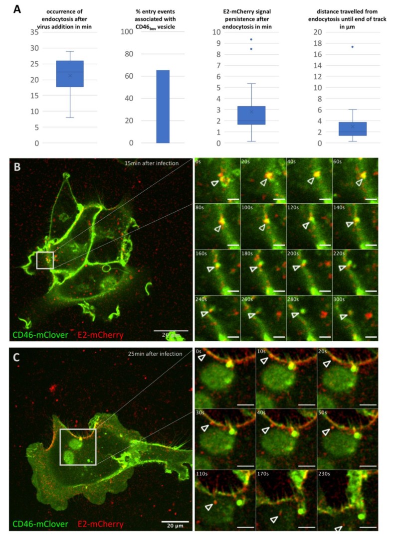
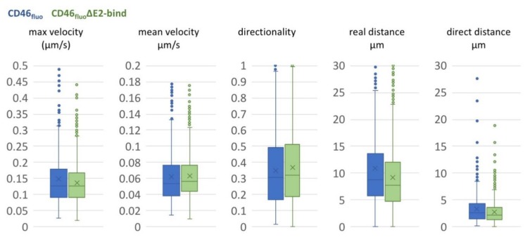
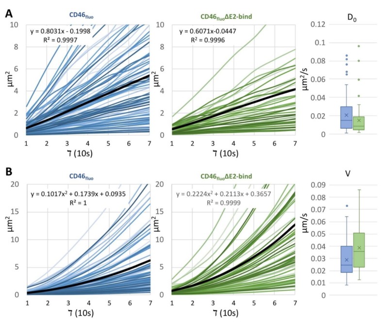
Virus attachment and entry is a complex interplay of viral and cellular interaction partners. Employing bovine viral diarrhea virus (BVDV) encoding an mCherry-E2 fusion protein (BVDVE2-mCherry), being the first genetically labelled member of the family Flaviviridae applicable for the analysis of virus particles, the early events of infection-attachment, particle surface transport, and endocytosis-were monitored to better understand the mechanisms underlying virus entry and their dependence on the virus receptor, bovine CD46. The analysis of 801 tracks on the surface of SK6 cells inducibly expressing fluorophore labelled bovine CD46 (CD46fluo) demonstrated the presence of directed, diffusive, and confined motion. 26 entry events could be identified, with the majority being associated with a CD46fluo positive structure during endocytosis and occurring more than 20 min after virus addition. Deletion of the CD46fluo E2 binding domain (CD46fluo∆E2bind) did not affect the types of motions observed on the cell surface but resulted in a decreased number of observable entry events (2 out of 1081 tracks). Mean squared displacement analysis revealed a significantly increased velocity of particle transport for directed motions on CD46fluo∆E2bind expressing cells in comparison to CD46fluo. These results indicate that the presence of bovine CD46 is only affecting the speed of directed transport, but otherwise not influencing BVDV cell surface motility. Instead, bovine CD46 seems to be an important factor during uptake, suggesting the presence of additional cellular proteins interacting with the virus which are able to support its transport on the virus surface.

Keywords: BVDV; CD46; Pestivirus; attachment; life cell imaging; surface transport.


Conflict of interest statement

The authors declare no conflict of interest.

Figures

Figure 1
Figure 1
Characterization of BVDVE2-mCherry entry into SK6 CD46fluo cells. (A) Characterization of 26 entry events of BVDVE2-mCherry in SK6 CD46fluo cells with regard to occurrence after virus addition, association with CD46fluo signal, intracellular persistence of the E2-mCherry signal and intracellular distance travelled. Outliers (Q3 + 1.5-times interquartile range) are indicated by dots. (B,C) Examples of entry events of BVDVE2-mCherry (red) into SK6 CD46bov (green) cells. The full field of view at the time of the start of acquisition (time after virus addition is specified in the top right corner) is shown and the area of interest is indicated by grey squares. Frames as depicted in the detail images were acquired every 10 s for up to 10 min after the start of acquisition. Times indicated in s refer to the start of acquisition. The length of the scale bar in the detail images in (B) = 2.5 µm and in (C) = 5 µm.
Figure 2
Figure 2
E2-mCherry signal development but not signal distribution is depending on the MOI. (A) Occurrence of E2-mCherry signal in min after addition of BVDVE2-mCherry at an MOI of 1 (blue) or 10 (green) to SK6 CD46fluo cells. SK6 CD46fluo cells were imaged recording one frame every 10 min at one z-level for 16 h. The mean is indicated by x and outliers (Q3 + 1.5-times interquartile range) are indicated by dots. (B) Distribution of E2-mCherry (red) and CD46fluo (green) signal 20 h after infection with BVDVE2-mCherry in SK6 CD46fluo cells. SK6 CD46fluo cells were imaged for 10 min recording 3 z-levels (0.8 µm) every 10 s (movie 6). Points of high E2-mCherry intensity that are colocalizing with CD46fluo are indicated by white circles.
Figure 3
Figure 3
Deletion of CCP-1 reduces CD46fluo effect of the susceptibility of SK6 cells to the level of GFP-expressing controls. Susceptibility of SK6 CD46fluo (blue), CD46fluo∆E2bind (green) and GFP-expressing (grey) SK6 cells to infection with BVDVE2-mCherry in % of MDBK cells (=100%) with and without the induction of expression by the addition of doxycycline (Doxy) (n = 6). Cells were infected with serial dilutions of BVDVE2-mCherry for 4 h. Subsequently, medium was exchanged to medium containing 1% carboxymethycellulose and E2-mCherry positive foci were detected 48 h after infection by fluorescence microscopy to determine the focus forming units (ffu)/mL. Susceptibility of a given cell line was calculated as the percentage of ffu/mL determined for MDBK cells, which was set to 100%.
Figure 4
Figure 4
Movements of BVDVE2-mCherry on the surface of SK6 CD46fluo (blue) or CD46fluo∆E2bind cells (green) are comparable. Box and Whisker blots of maximum and mean velocity, directionality, real distance and direct distance of particles tracked on the surface of SK6 CD46fluo (blue, n = 801) or CD46fluo∆E2bind cells (green, n = 1081), respectively. The mean is indicated by x and outliers (Q3 + 1.5-times interquartile range) are indicated by dots.
Figure 5
Figure 5
The average velocity of directed motion of BVDVE2-mCherry particles is increased on the surface of SK6 CD46fluo∆E2bind cells. MSD analysis of particle movement on SK6 CD46fluo and CD46fluo∆E2bind cells. (A) Slopes of tracks on SK6 CD46fluo (blue) and CD46fluo∆E2bind (green) cells displaying a linear increase and distribution of the calculated diffusion coefficient D0 depicted as box and whisker blot. The mean is indicated by x and outliers (Q3 + 1.5 times interquartile range) are depicted as dots. Average slopes are depicted as thick black lines. (B) Slopes of tracks on SK6 CD46fluo (blue) and CD46fluo∆E2bind (green) cells displaying an exponential increase and distribution of the calculated particle velocities V, depicted as box and whisker blot. The mean is indicated by x and outliers (Q3 + 1.5 times interquartile range) are depicted as dots. Average slopes are depicted as thick black lines.

Similar articles

Cited by

References

    1. Burckhardt C.J., Greber U.F. Virus movements on the plasma membrane support infection and transmission between cells. PLoS Pathog. 2009;5:e1000621. doi: 10.1371/journal.ppat.1000621. - DOI - PMC - PubMed
    1. Lehmann M.J., Sherer N.M., Marks C.B., Pypaert M., Mothes W. Actin- and myosin-driven movement of viruses along filopodia precedes their entry into cells. J. Cell Biol. 2005;170:317–325. doi: 10.1083/jcb.200503059. - DOI - PMC - PubMed
    1. Oh M.-J., Akhtar J., Desai P., Shukla D. A role for heparan sulfate in viral surfing. Biochem. Biophys. Res. Commun. 2010;391:176–181. doi: 10.1016/j.bbrc.2009.11.027. - DOI - PMC - PubMed
    1. Schelhaas M., Shah B., Holzer M., Blattmann P., Kühling L., Day P.M., Schiller J.T., Helenius A. Entry of human papillomavirus type 16 by actin-dependent, clathrin- and lipid raft-independent endocytosis. PLoS Pathog. 2012;8:e1002657. doi: 10.1371/journal.ppat.1002657. - DOI - PMC - PubMed
    1. Ayala-Nuñez N.V., Wilschut J., Smit J.M. Monitoring virus entry into living cells using DiD-labeled dengue virus particles. Methods. 2011;55:137–143. doi: 10.1016/j.ymeth.2011.07.009. - DOI - PubMed

Publication types

LinkOut - more resources