Review
. 2020 Jan 17;12(1):117.
doi: 10.3390/v12010117.

Microtubules in Influenza Virus Entry and Egress

Affiliations
Review

Microtubules in Influenza Virus Entry and Egress

Caitlin Simpson et al. Viruses. .

Abstract

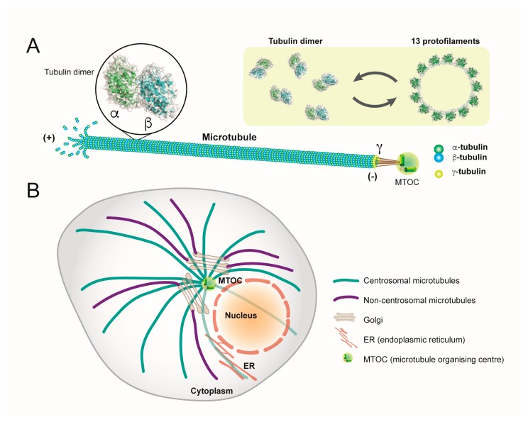
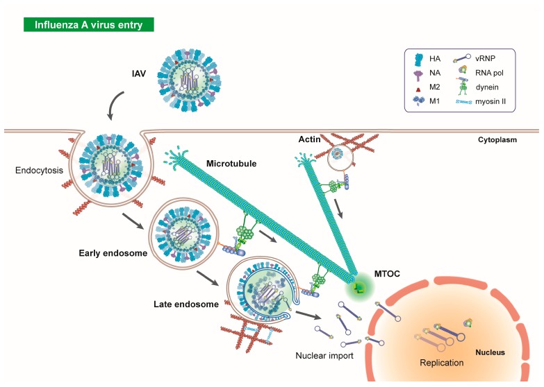
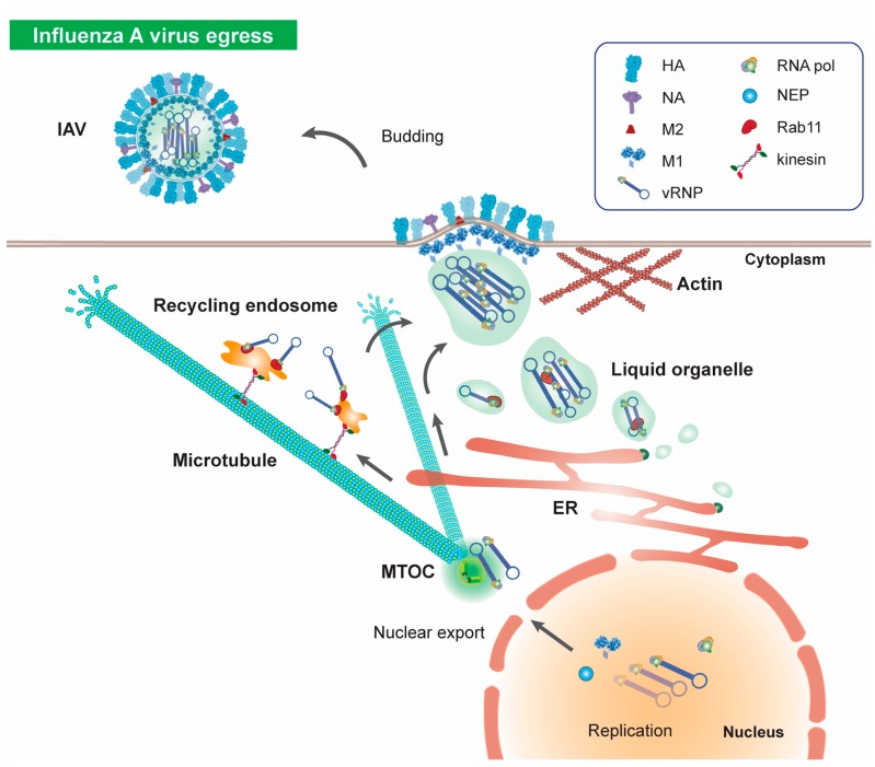
Influenza viruses are respiratory pathogens that represent a significant threat to public health, despite the large-scale implementation of vaccination programs. It is necessary to understand the detailed and complex interactions between influenza virus and its host cells in order to identify successful strategies for therapeutic intervention. During viral entry, the cellular microenvironment presents invading pathogens with a series of obstacles that must be overcome to infect permissive cells. Influenza hijacks numerous host cell proteins and associated biological pathways during its journey into the cell, responding to environmental cues in order to successfully replicate. The cellular cytoskeleton and its constituent microtubules represent a heavily exploited network during viral infection. Cytoskeletal filaments provide a dynamic scaffold for subcellular viral trafficking, as well as virus-host interactions with cellular machineries that are essential for efficient uncoating, replication, and egress. In addition, influenza virus infection results in structural changes in the microtubule network, which itself has consequences for viral replication. Microtubules, their functional roles in normal cell biology, and their exploitation by influenza viruses will be the focus of this review.

Keywords: aggresome processing; cytoskeleton; endocytosis; histone deacetylase; infection biology; influenza virus; microtubules; uncoating.


Conflict of interest statement

The authors declare no conflict of interest.

Figures

Figure 1
Figure 1
Structure and organisation of microtubules. (A) Microtubule filaments are comprised of multiple dimeric complexes of α- and β-tubulin, assembled around a hollow core. Thirteen protofilaments assemble to form a microtubule. Microtubules are anchored at their minus ends at MTOCs, which is mediated by γ-tubulin. (B) Microtubules form dynamic networks in the cytoplasm which are stably anchored at MTOCs, including the centrosome and Golgi apparatus. Three-dimensional structural data: PDB ID tubulin dimer (1TUB).
Figure 2
Figure 2
Influenza A virus endocytosis and early trafficking through the cell. IAV is a single-stranded negative sense RNA virus, belonging to the Orthomyxoviridae family. Viral particles are composed of an outer envelope containing the glycoproteins hemagglutinin (HA) and neuraminidase (NA) and M2 ion channels. An M1 shell constitutes the viral shell, within which are 8 viral gene segments each in association with nucleoprotein (NP) and an RNA polymerase. The influenza polymerase itself is formed from three subunits; PA, PB1 and PB2. Following attachment of IAV to permissive cells via sialylated cell-surface receptors, the virus is endocytosed via clathrin-mediated endocytosis and macropinocytosis. After initial association with the actin-myosin network, early endosomes containing IAV virions interact with microtubules via dynein motor proteins for retrograde traffic towards the MTOC, in close proximity to the cellular nucleus. Upon reaching the perinuclear region, IAVs undergo low-pH mediated fusion with the late endosomal membrane. M1 shell uncoating is dependent on microtubules, actin, and the motors dynein and myosin II. Release of vRNPs into the cytosol and uptake into the nucleus precedes viral genome replication. Three-dimensional structural data: PDB ID HA (2IBX); NA (6CRD); M2 (3BKD); M1 (1EA3) [65].
Figure 3
Figure 3
Influenza A virus egress. Following replication of viral RNA, newly synthesised vRNPs are exported from the nucleus and accumulate at the MTOC, before trafficking towards the cellular periphery in a microtubule dependent manner for assembly and budding. Influenza viruses utilise components of the endocytic recycling and secretory pathways for apical transport; associations between Rab11-positive recycling endocytic vesicles and influenza viruses allow viral traffic along microtubules. In addition, vRNPs induce formation of liquid organelles which associate with Rab11 for vRNP traffic via the secretory pathway. Following microtubule-dependent anterograde traffic to the cellular periphery, vRNPs assemble to form new virions and bud from the cell surface to trigger secondary infection in permissive cells. Three-dimensional structural data: PDB ID HA (2IBX); NA (6CRD); M2 (3BKD); M1 (1EA3) [65].

Similar articles

Cited by

References

    1. Bedi S., Ono A. Friend or Foe: The Role of the Cytoskeleton in Influenza A Virus Assembly. Viruses. 2019;11:46. doi: 10.3390/v11010046. - DOI - PMC - PubMed
    1. Muroyama A., Lechler T. Microtubule organization, dynamics and functions in differentiated cells. Development. 2017;144:3012–3021. doi: 10.1242/dev.153171. - DOI - PMC - PubMed
    1. Wickstead B., Gull K. The evolution of the cytoskeleton. J. Cell Biol. 2011;194:513–525. doi: 10.1083/jcb.201102065. - DOI - PMC - PubMed
    1. Akhmanova A., Steinmetz M.O. Control of microtubule organization and dynamics: Two ends in the limelight. Nat. Rev. Mol. Cell Biol. 2015;16:711–726. doi: 10.1038/nrm4084. - DOI - PubMed
    1. Cai D., McEwen D.P., Martens J.R., Meyhofer E., Verhey K.J. Single molecule imaging reveals differences in microtubule track selection between Kinesin motors. PLoS Biol. 2009;7:e1000216. doi: 10.1371/journal.pbio.1000216. - DOI - PMC - PubMed

LinkOut - more resources