Review
. 2020 Jan 17;9(1):257.
doi: 10.3390/jcm9010257.

Regulation of BDNF-TrkB Signaling and Potential Therapeutic Strategies for Parkinson's Disease

Affiliations
Review

Regulation of BDNF-TrkB Signaling and Potential Therapeutic Strategies for Parkinson's Disease

Wook Jin. J Clin Med. .

Abstract

Brain-derived neurotrophic factor (BDNF) and its receptor tropomyosin-related kinase receptor type B (TrkB) are widely distributed in multiple regions of the human brain. Specifically, BDNF/TrkB is highly expressed and activated in the dopaminergic neurons of the substantia nigra and plays a critical role in neurophysiological processes, including neuro-protection and maturation and maintenance of neurons. The activation as well as dysfunction of the BDNF-TrkB pathway are associated with neurodegenerative diseases. The expression of BDNF/TrkB in the substantia nigra is significantly reduced in Parkinson's Disease (PD) patients. This review summarizes recent progress in the understanding of the cellular and molecular roles of BNDF/TrkB signaling and its isoform, TrkB.T1, in Parkinson's disease. We have also discussed the effects of current therapies on BDNF/TrkB signaling in Parkinson's disease patients and the mechanisms underlying the mutation-mediated acquisition of resistance to therapies for Parkinson's disease.

Keywords: BDNF; Parkinson’s disease; TrkB; TrkB isoform; neuronal degeneration.


Conflict of interest statement

The author declares no conflict of interest.

Figures

Figure 1
Figure 1
Brain-derived neurotrophic factor (BDNF)/tropomyosin-related kinase receptor type B (BDNF/TrkB) signaling supports neuronal survival, plasticity, differentiation, and growth via activation of several functional downstream cascades. Binding BDNF to TrkB as its specific receptor leads to homodimerization and triggers activation of adaptor proteins such as polypyrimidine tract-binding protein (PTB) and Src homology domain 2 (SH2). Subsequently, activated adaptor proteins lead to activation of phosphoinositide 3-kinases (PI3K)-AKT (PI3K-AKT), Ras-mitogen-activated protein kinase (Ras-MAPK), and phospholipase Cγ1 (PLC-γ1)-protein kinase C (PKC) signaling pathway.
Figure 2
Figure 2
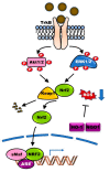
BDNF-TrkB signaling protects neurons from reactive oxygen species (ROS)-induced cell death. BDNF-TrkB signaling leads to the activation of phosphoinositide 3-kinases (PI3K)-AKT (PI3K-AKT) and Ras-mitogen-activated protein kinase (Ras-MAPK) pathways. Activated PI3K-AKT and Ras-MAPK pathways trigger dissociation of nuclear factor erythroid 2-related factor 2 (Nrf2)-Keap1 complex and then induces nuclear translocation of Nrf2. Finally, the binding of Nrf2 to antioxidant response element (ARE) in target genes leads to the expression of antioxidant enzymes, including heme oxygenase-I (HO-1) and, subsequently, involved in protection from ROS-mediated neuronal cell death of Parkinson’s Disease (PD).
Figure 3
Figure 3
The generation of endoplasmic reticulum (ER) stress by ROS and accumulation of α-syn aggregates disrupts BDNF-TrkB-mediated neuronal protection. Accumulation or mutation of α-syn in PD reduces BDNF or TrkB expression through TrkB ubiquitination, and its reduction of BDNF-TrkB signaling leads to the generation of ER stress by induction of ROS or accumulation of α-syn. ER stress triggers the misfolding of proteins, leading to the accumulation of protein aggregates. Accumulation of protein aggregates activates protein kinase RNA-like ER kinase (PERK), activating transcription factor (ATF6α), and inositol-requiring protein (IRE1α) as three main transmembrane proteins in the ER. The activation of PERK, which induces eukaryotic Initiation Factor 2α (eIF2α) phosphorylation, inhibits protein translation and increases ATF4, which is involved in apoptosis through induction of the CCAAT-enhancer-binding protein (C/EBP) homologous protein (CHOP) known as growth arrest and DNA damage-inducible protein. Also, activated IRE1α induced neuronal apoptosis by induction of c-Jun N-terminal kinase (JNK) phosphorylation, or by increasing inflammatory response inducting IRE1α-dependent messenger RNA decay (RIDD). Additionally, the production of ATF6 fragment (ATF6f) by cleavage of ATF6 in the Golgi apparatus is involved in the development of PD by inducing apoptosis of neuronal cells.
Figure 4
Figure 4
Schematic representation of TrkB full length and truncated isoforms. The domain of TrkB exhibited as filled boxes. TrkB-N, TrkB-N-T-TK, TrkB-N-T-Shc, and TrkB-N-T1 have a lack of N-terminal signal sequence, leucine-rich repeat N-terminal domain (LRRNT), leucine-rich repeat region 1 (LRR1), and LRR2 domain of TrkB. Also, TrkB-L1 and TrkB-L0 lacked the first two or three or all three of LRRs in the extracellular domain of TrkB, respectively. Additionally, TrkB.T1 and TrkB.T2, TrkB-T-Shc, TrkB-T’ lack c-terminal region of TrkB, including tyrosine kinase domain. LRRCT, leucine-rich repeat C-terminal domain; Ig-like C2, immunoglobulin-C2-set domain; TM, transmembrane domain.

Similar articles

Cited by

References

    1. Arevalo J.C., Wu S.H. Neurotrophin signaling: Many exciting surprises! Cell. Mol. Life Sci. 2006;63:1523–1537. doi: 10.1007/s00018-006-6010-1. - DOI - PMC - PubMed
    1. Zaccaro M.C., Ivanisevic L., Perez P., Meakin S.O., Saragovi H.U. p75 co-receptors regulate ligand-dependent and ligand-independent Trk receptor activation, in part by altering Trk docking subdomains. J. Biol. Chem. 2001;276:31023–31029. doi: 10.1074/jbc.M104630200. - DOI - PubMed
    1. Castren E., Zafra F., Thoenen H., Lindholm D. Light Regulates Expression of Brain-Derived Neurotrophic Factor Messenger-Rna in Rat Visual-Cortex. Proc. Natl. Acad. Sci. USA. 1992;89:9444–9448. doi: 10.1073/pnas.89.20.9444. - DOI - PMC - PubMed
    1. Bracken B.K., Turrigiano G.G. Experience-Dependent Regulation of TrkB Isoforms in Rodent Visual Cortex. Dev. Neurobiol. 2009;69:267–278. doi: 10.1002/dneu.20701. - DOI - PMC - PubMed
    1. Patterson S.L., Abel T., Deuel T.A.S., MArtin K.C., Rose J.C., Kandel E.R. Recombinant BDNF rescues deficits in basal synaptic transmission and hippocampal LTP in BDNF knockout mice. Neuron. 1996;16:1137–1145. doi: 10.1016/S0896-6273(00)80140-3. - DOI - PubMed

LinkOut - more resources