Review
. 2020 Jan 17;21(2):626.
doi: 10.3390/ijms21020626.

Age-Associated TET2 Mutations: Common Drivers of Myeloid Dysfunction, Cancer and Cardiovascular Disease

Affiliations
Review

Age-Associated TET2 Mutations: Common Drivers of Myeloid Dysfunction, Cancer and Cardiovascular Disease

Christina K Ferrone et al. Int J Mol Sci. .

Abstract

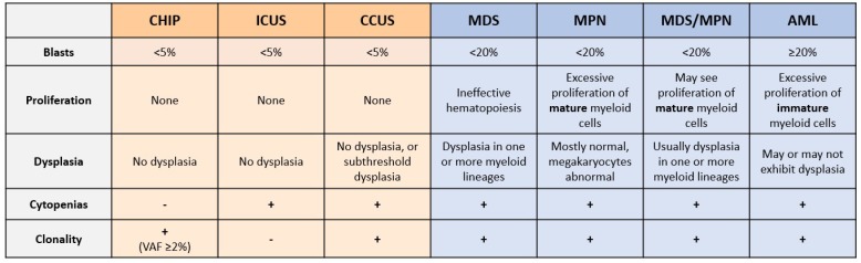
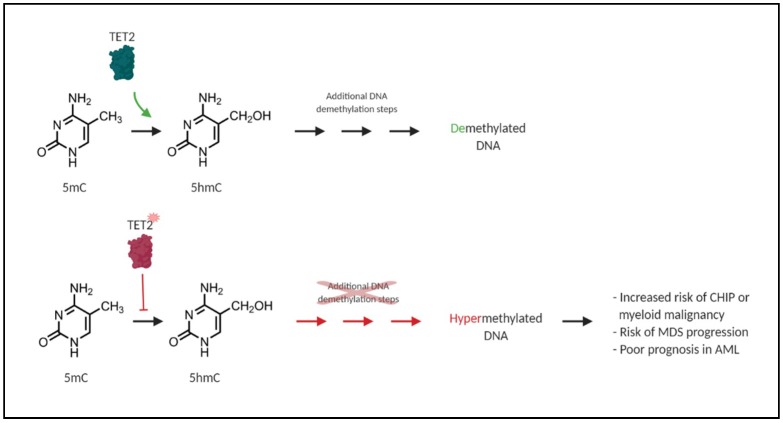
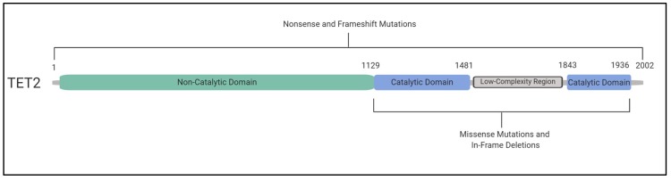
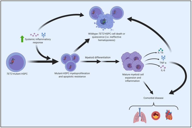
Acquired, inactivating mutations in Tet methylcytosine dioxygenase 2 (TET2) are detected in peripheral blood cells of a remarkable 5%-10% of adults greater than 65 years of age. They impart a hematopoietic stem cell advantage and resultant clonal hematopoiesis of indeterminate potential (CHIP) with skewed myelomonocytic differentiation. CHIP is associated with an overall increased risk of transformation to a hematological malignancy, especially myeloproliferative and myelodysplastic neoplasms (MPN, MDS) and acute myeloid leukemia (AML), of approximately 0.5% to 1% per year. However, it is becoming increasingly possible to identify individuals at greatest risk, based on CHIP mutational characteristics. CHIP, and particularly TET2-mutant CHIP, is also a novel, significant risk factor for cardiovascular diseases, related in part to hyper-inflammatory, progeny macrophages carrying TET2 mutations. Therefore, somatic TET2 mutations contribute to myeloid expansion and innate immune dysregulation with age and contribute to prevalent diseases in the developed world-cancer and cardiovascular disease. Herein, we describe the impact of detecting TET2 mutations in the clinical setting. We also present the rationale and promise for targeting TET2-mutant and other CHIP clones, and their inflammatory environment, as potential means of lessening risk of myeloid cancer development and dampening CHIP-comorbid inflammatory diseases.

Keywords: NGS; TET2; aging; cancer progression; clinical detection; clonal hematopoiesis; comorbid disease; driver mutations; inflammation; targeting TET2 therapeutically.


Conflict of interest statement

The authors declare no conflict of interest.

Figures

Figure 1
Figure 1
Summary of current pre-neoplastic states and myeloid neoplasms. Pre-neoplastic states (in orange: clonal hematopoiesis of indeterminate potential, CHIP; idiopathic cytopenia of undetermined significance, ICUS; and clonal cytopenias of undetermined significance; CCUS) are classified primarily based on the presence of clonality (acquired DNA variants or chromosomal aberrations), and on the presence of peripheral cytopenias [13,14]. The main categories of myeloid neoplasm (blue) are primarily classified based on blast percentage (morphologically primitive cells, including HSPCs), by the type of cells that proliferate aberrantly, and by the morphology of the myeloid cells [15]. VAF, variant allele frequency; MDS, myelodysplastic syndromes; MPN, myeloproliferative neoplasms; MDS/MPN, overlapping MDS and MPN; AML, acute myeloid leukemia.
Figure 2
Figure 2
Loss-of-function mutations in TET2 result in DNA hypermethylation. TET2 is an alpha-ketoglutarate- and Fe2+-dependent dioxygenase (α-KGDD) that catalyzes the oxidation of 5-methylcytosine (5mC) to 5-hydroxymethylcytosine (5hmC). This is a required step in proper DNA repair and DNA demethylation (green). Loss-of-function mutations in TET2 in CHIP and myeloid malignancies disrupt this oxidation step and result in a general DNA hypermethylation phenotype (red) and aberrant HSPC self-renewal, which is associated with an increased risk of CHIP, myeloid malignancy, MDS progression, and poor prognosis in AML [35].
Figure 3
Figure 3
Inactivating TET2 mutations occur throughout the coding region. The C-terminal catalytic domain of TET2 occurs approximately between amino acid residues 1129 and 1936, including a double-stranded β helix and cysteine-rich domain, binding sites for Fe(II) and α-ketoglutarate, as well as a low-complexity linker region. Together, these regions allow the catalytic domain of TET2 to bind to DNA, allowing the oxidation of 5mC to 5hmC [40]. Missense mutations and in-frame deletions occur in the catalytic domain of the protein (blue). Nonsense and frameshift mutations can occur throughout the entire coding region. However, they occur most frequently in the non-catalytic domain (green). Mutations in TET2 are generally inactivating regardless of their nature or location in the coding region of the gene [8,40,41].
Figure 4
Figure 4
Molecular pathogenesis of TET2-mutated HSPC. Exposure to upregulated inflammatory cytokines (due to systemic inflammation or local alterations) advantages TET2-mutant HSPCs, fostering a phenotype characterized by increased proliferation and a resistance to apoptosis [69,70,71]. TET2-mutant HSPCs and the abnormal immune microenvironment of the bone marrow can cause premature death or quiescence of wildtype (i.e., normal) HSPCs, consequently manifesting as ineffective and clonal hematopoiesis. The increased proliferation associated with TET2-mutant HSPCs leads to the expansion of TET2-clonal populations in the bone marrow and/or peripheral blood. Mature myeloid cells (e.g., macrophages) derived from TET2-mutant HSPCs demonstrate a hyper-inflammatory phenotype that contributes to the pathogenesis of comorbidities, with a notable causal role in cardiovascular disease [57,61,67].

Similar articles

Cited by

References

    1. Arber D.A., Orazi A., Hasserjian R., Thiele J., Borowitz M.J., Le Beau M.M., Bloomfield C.D., Cazzola M., Vardiman J.W. The 2016 revision to the World Health Organization classification of myeloid neoplasms and acute leukemia. Blood. 2016;127:2391–2406. doi: 10.1182/blood-2016-03-643544. - DOI - PubMed
    1. Barbui T., Thiele J., Gisslinger H., Kvasnicka H.M., Vannucchi A.M., Guglielmelli P., Orazi A., Tefferi A. The 2016 WHO classification and diagnostic criteria for myeloproliferative neoplasms: Document summary and in-depth discussion. Blood Cancer J. 2018;8:15. doi: 10.1038/s41408-018-0054-y. - DOI - PMC - PubMed
    1. Haferlach T., Nagata Y., Grossmann V., Okuno Y., Bacher U., Nagae G., Schnittger S., Sanada M., Kon A., Alpermann T., et al. Landscape of genetic lesions in 944 patients with myelodysplastic syndromes. Leukemia. 2014;28:241–247. doi: 10.1038/leu.2013.336. - DOI - PMC - PubMed
    1. Mardis E.R., Ding L., Dooling D.J., Larson D.E., McLellan M.D., Chen K., Koboldt D.C., Fulton R.S., Delehaunty K.D., McGrath S.D., et al. Recurring mutations found by sequencing an acute myeloid leukemia genome. N. Engl. J. Med. 2009;361:1058–1066. doi: 10.1056/NEJMoa0903840. - DOI - PMC - PubMed
    1. Papaemmanuil E., Gerstung M., Malcovati L., Tauro S., Gundem G., Loo P.V., Yoon C.J., Ellis P., Wedge D.C., Pellagatti A., et al. Clinical and biological implications of driver mutations in myelodysplastic syndromes. Blood. 2013;122:3616–3627. doi: 10.1182/blood-2013-08-518886. - DOI - PMC - PubMed