. 2020 Jan 18;9(1):263.
doi: 10.3390/jcm9010263.

Ringer's Lactate Prevents Early Organ Failure by Providing Extracellular Calcium

Affiliations

Ringer's Lactate Prevents Early Organ Failure by Providing Extracellular Calcium

Biswajit Khatua et al. J Clin Med. .

Abstract

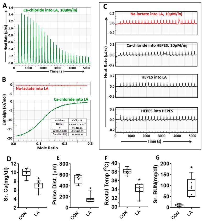
Objective: Ringer's lactate may improve early systemic inflammation during critical illnesses like severe acute pancreatitis, which are associated with hypocalcemia. Ringer's lactate is buffered and contains lactate and calcium. We, thus analyzed extracellular calcium or lactate's effects on the mechanisms, intermediary markers, and organ failure in models mimicking human disease with nonesterified fatty acid (NEFA) elevation.

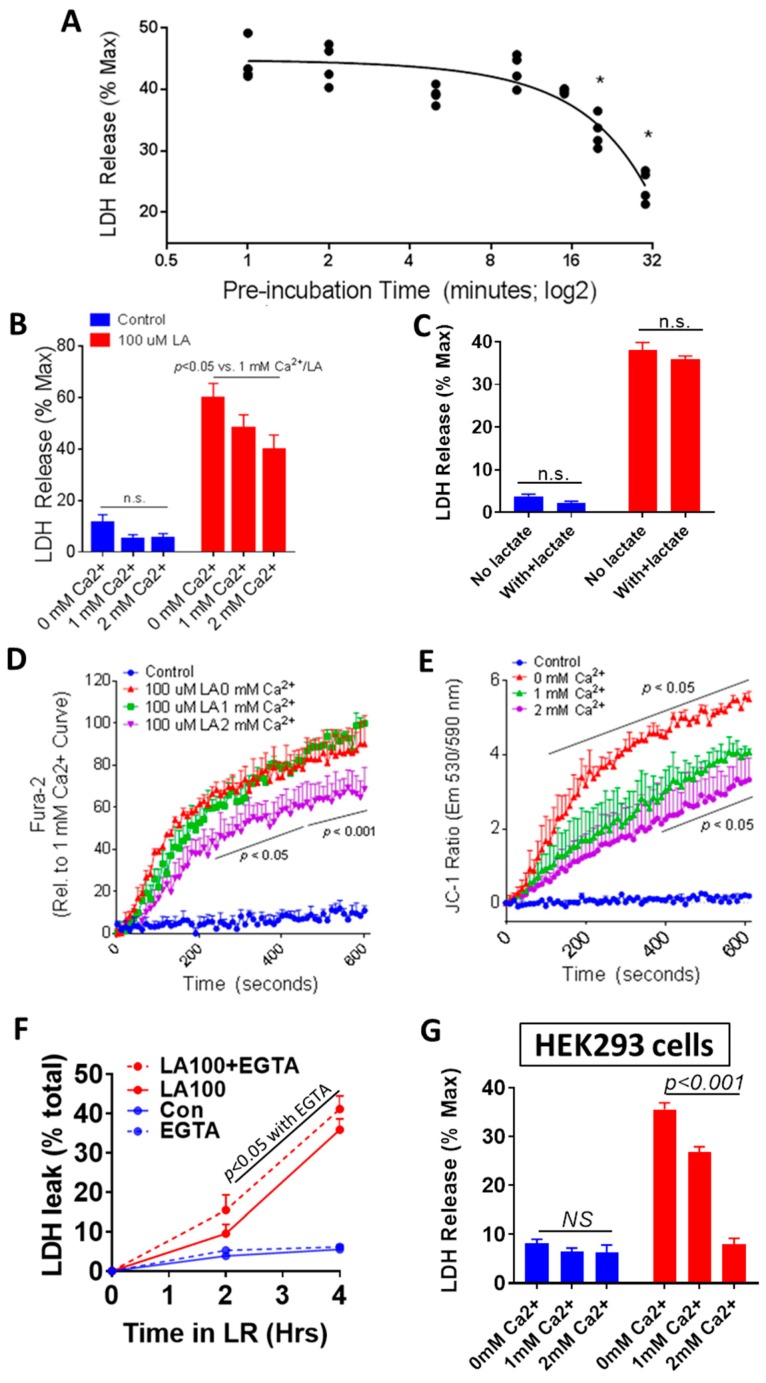
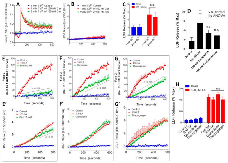
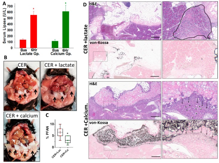
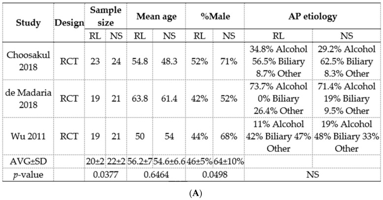
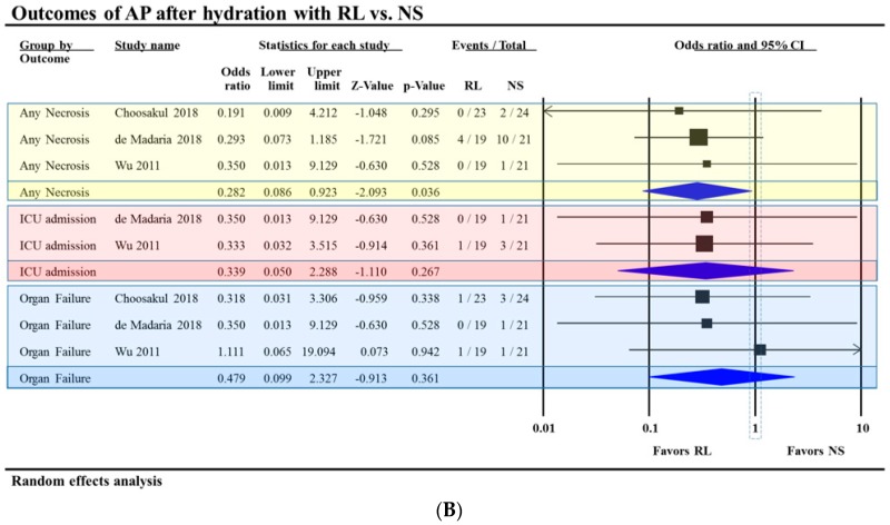
Methods: Meta-analyses and experimental studies were performed. Experimentally, extracellular calcium and lactate were compared in their interaction with linoleic acid (LA; a NEFA increased in human severe pancreatitis), and its subsequent effects on mitochondrial depolarization and cytosolic calcium signaling resulting in cell injury. In vivo, the effect of LA was studied on organ failure, along with the effect of calcium or lactate (pH 7.4) on severe acute pancreatitis-associated organ failure. A meta-analysis of human randomized control trials comparing Ringer's lactate to normal saline was done, focusing on necrosis and organ failure.

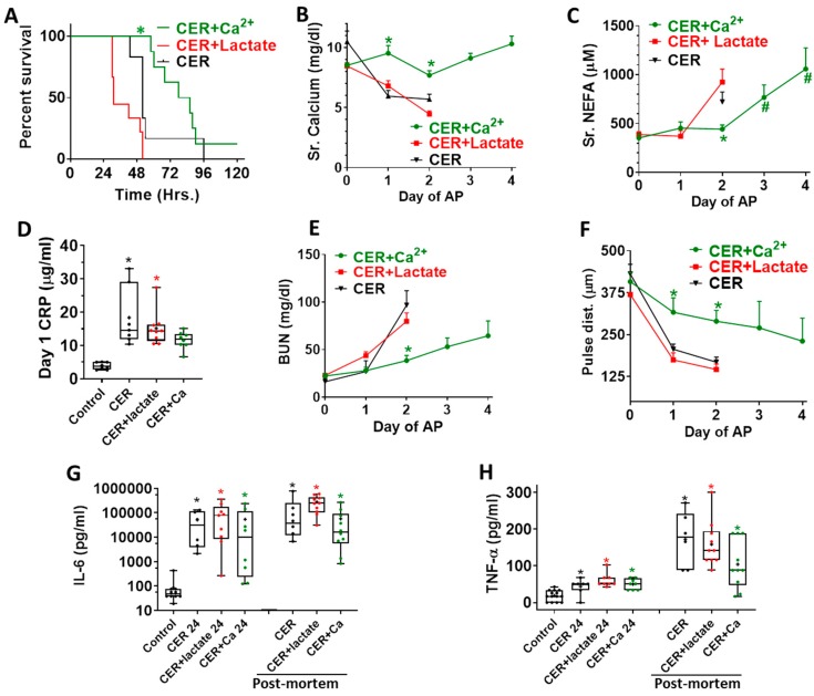
Results: Calcium reacted ionically with LA and reduced lipotoxic necrosis. In vivo, LA induced organ failure and hypocalcemia. During severe pancreatitis, calcium supplementation in saline pH 7.4, unlike lactate, prevented hypocalcemia, increased NEFA saponification, reduced circulating NEFA and C-reactive protein , reduced pancreatic necrosis adjacent to fat necrosis, and normalized shock (carotid pulse distension) and blood urea nitrogen elevation on day 1. This, however, did not prevent the later increase in serum NEFA which caused delayed organ failure. Meta-analysis showed Ringer's lactate reduced necrosis, but not organ failure, compared with normal saline.

Conclusion: Hypocalcemia occurs due to excess NEFA binding calcium during a critical illness. Ringer's lactate's early benefits in systemic inflammation are by the calcium it provides reacting ionically with NEFA. This, however, does not prevent later organ failure from sustained NEFA generation. Future studies comparing calcium supplemented saline resuscitation to Ringer's lactate may provide insights to this pathophysiology.

Keywords: CRP; Ringer’s lactate; calcium; inflammation; isothermal titration calorimetry; lipolysis; mitochondrial depolarization; organ failure; pancreatitis; saponification.


Conflict of interest statement

The authors have declared that no conflicts of interest exist.

Figures

Figure 1
Figure 1
(AC): Isothermal titration calorimetry showing Linoleic acid (LA) reacts with calcium but not lactate. (A) Thermogram showing heat rate of injection of calcium chloride (10 μM/injection) into 1 mM Linoleic acid (LA). Both were dissolved in 10 mM HEPES (pH 7.4) (B) Enthalpograms of injection of sodium lactate (Na-lactate, red line on top) or calcium chloride (CaCl2; green line below) into linoleic acid. The various thermodynamic variables (Mean ± SD (standard deviation)) from three different experiments are mentioned in the table adjacent to the thermogram. (C) Thermogram showing heat rate of injection of sodium lactate (top, red line; 10 μM/injection) and other control experimental conditions below this (black lines). (DG): Effects of intraperitoneal administration of linoleic acid (LA, 0.1% body weight) to CD-1 mice on their serum calcium (D), carotid pulse distension (pulse dist.) (E), rectal temperature (F), and blood urea nitrogen (Sr. BUN) (G). The vitals were measured at 40 h, and blood parameters at necropsy.* indicates a p-value of <0.05 on Mann-Whitney test.
Figure 2
Figure 2
Extracellular calcium reduces LA-induced acinar cell injury in vitro. (A) Time course of the effect of different durations of preincubating LA with calcium at 37 °C prior to exposure to primary pancreatic acini, showing its impact on LDH release into the medium of the acini. (B) Extracellular calcium dose-dependently reduces 100 μM LA-induced acinar LDH release over 4 h (red bars) without affecting control cell viability (blue bars). (C) Supplementation with 10 mM sodium lactate had no effect on either baseline cell viability or 100 μM LA-induced acinar cell injury. Effect of different doses of extracellular calcium (0 mM red line, 1 mM green line, 2 mM purple line) on 100 μM LA-induced intracellular calcium elevation (D) and mitochondrial depolarization (E). (F) Chelation of extracellular calcium (1 mM) with EGTA (1 mM) to nominally absent levels increased LA-induced acinar cell injury (dotted red line) at 2 and 4 h. (G): Extracellular calcium dose-dependently reduces 100 μM LA-induced LDH release from HEK293 cells over 4 h (red bars) without affecting control cell viability (blue bars).
Figure 3
Figure 3
Effect of altering calcium on caerulein-induced (AD), and changing Cai on LA-induced (EH) acinar cell injury. (A) Extracellular calcium (1, 2 mM) had no effect on intracellular calcium peak and plateau levels induced by 100 nM caerulein and (B) did not modulate the lack of mitochondrial depolarization with caerulein treatment. (C) Extracellular calcium did not reduce caerulein-induced acinar cell injury, while (D) intracellular calcium chelation with BAPTA-AM (50 μM) or ryanodine receptor antagonization with dantrolene (100 μM) caused caerulein-induced injury to be nonsignificant versus control. (E) BAPTA-AM (50 μM), (F) Dantrolene (100 μM), and (G) thapsigargin (1 μM) all reduced intracellular calcium elevation after 100 μM LA treatment, while BAPTA-AM (E’) partially limiting mitochondrial depolarization Dantrolene (F’) and thapsigargin (G’) had no effect on mitochondrial depolarization. (H): Despite effects on calcium elevation, BAPTA-AM, Dantrolene, and Thapsigargin did not significantly affect LA-induced injury in acinar cells, as measured by LDH release.
Figure 4
Figure 4
Comparison of the therapeutic calcium and lactate on fat necrosis during severe experimental pancreatitis: (A) serum lipase levels as measured under basal (Bas) conditions before induction of pancreatitis and after 6 h of pancreatitis (6 H), following which either Sodium lactate or Calcium chloride were given intraperitoneally, as described in the methods section. (B) Gross appearance of the peritoneal cavity at necropsy of mice with caerulein pancreatitis alone (CER), caerulein pancreatitis treated with sodium lactate (CER + lactate), or caerulein pancreatitis treated with calcium chloride (CER + calcium). Note the much larger extent of white- colored saponification of fat necrosis in the calcium-treated group (black arrows). (C) Box plots comparing the extent of peri-fat acinar necrosis (% PFAN) measured as a percentage of total pancreatic parenchymal area in the CER + lactate (CER + Lac)- and CER + calclium (CER + Ca)-treated groups. * indicates a p < 0.05. (D) Histologic images of the pancreas and adjacent fat necrosis stained with hematoxylin and eosin (H&E), and von-Kossa for calcium, in the CER + lactate and CER + calcium groups. To the right are zoomed-in images of the inset boxes highlighting the peri-fat acinar necrosis adjacent to the fat necrosis. The scale bar is 200 μm. Fat necrosis resulting in binding of calcium to the NEFA generated is seen as the pinkish stain in adipose tissue on H&E. Note the higher intensity of this in the CER + calcium group using both types of staining, especially von-Kossa, which extends into the parenchyma as a sharply demarcated line (black arrows). The peri-fat acinar necrosis is seen as loss of acinar cell outline and diffuse pale pinkish appearance of acini replacing the normal intensely pink zymogen and blue-colored basal cytoplasm. In the CER + calcium group, which survive longer, the prolonged caerulein stimulation results in acinar ductal metaplasia-like appearance with large central lumens replacing the zymogen (shown as *).
Figure 5
Figure 5
Effect of no treatment (black), calcium (green), or lactate treatment (red) on parameters of systemic injury and inflammation: (A) Kaplan–Meyer survival curve of the various groups with pancreatitis * indicates a p < 0.05 in the CER + calcium (CER + Ca2+)-treated group vs. other groups. (B) serum calcium and (C) Serum NEFA, as measured at each day of pancreatitis. * indicates a p < 0.05 in the CER + calcium treated groups vs. other groups on ANOVA. # indicates a significant difference from baseline (day 0). (D) Blood C-reactive protein (CRP) after 24 h of pancreatitis. * indicates a significant increase in the group on ANOVA vs. control mice without pancreatitis. (E) Blood urea nitrogen (BUN) and (F) Carotid artery pulse distention (pulse dist.), as measured on each day of pancreatitis. * indicates a p < 0.05 in the cer + calcium-treated groups vs. other groups on ANOVA. Interleukin 6 (IL-6) (G) and tumor necrosis factor (TNF-α) (H) blood levels, one day after pancreatitis initiation (each title followed by 24), and on the post-mortem blood samples. * indicates a significant increase on ANOVA for that time point vs. control.
Figure 6
Figure 6
Meta-analysis of three randomized controlled trials comparing Ringer’s lactate to normal saline. (A) Study characteristics of those included in the meta-analysis. RL: Ringer’s lactate. NS: Normal Saline. AP: Acute pancreatitis. AVG: Average. SD: Standard deviation. NS: Not significant. (B) Outcomes of acute pancreatitis after hydration with Ringer’s lactate vs. normal saline. AP: Acute pancreatitis. RL: Ringer’s lactate NS: Normal Saline.
Figure 6
Figure 6
Meta-analysis of three randomized controlled trials comparing Ringer’s lactate to normal saline. (A) Study characteristics of those included in the meta-analysis. RL: Ringer’s lactate. NS: Normal Saline. AP: Acute pancreatitis. AVG: Average. SD: Standard deviation. NS: Not significant. (B) Outcomes of acute pancreatitis after hydration with Ringer’s lactate vs. normal saline. AP: Acute pancreatitis. RL: Ringer’s lactate NS: Normal Saline.

References

    1. Banks P.A., Bollen T.L., Dervenis C., Gooszen H.G., Johnson C.D., Sarr M.G., Tsiotos G.G., Vege S.S. Classification of acute pancreatitis—2012: Revision of the Atlanta classification and definitions by international consensus. Gut. 2013;62:102–111. doi: 10.1136/gutjnl-2012-302779. - DOI - PubMed
    1. Mounzer R., Langmead C.J., Wu B.U., Evans A.C., Bishehsari F., Muddana V., Singh V.K., Slivka A., Whitcomb D.C., Yadav D., et al. Comparison of existing clinical scoring systems to predict persistent organ failure in patients with acute pancreatitis. Gastroenterology. 2012;142:1476–1482. doi: 10.1053/j.gastro.2012.03.005. - DOI - PubMed
    1. Wang B., Gong Y., Ying B., Cheng B. Association of Initial Serum Total Calcium Concentration with Mortality in Critical Illness. Biomed. Res. Int. 2018;2018:7648506. doi: 10.1155/2018/7648506. - DOI - PMC - PubMed
    1. Wang B., Li D., Gong Y., Ying B., Cheng B. Association of serum total and ionized calcium with all-cause mortality incritically ill patients with acute kidney injury. Clin. Chim. Acta. 2019;494:94–99. doi: 10.1016/j.cca.2019.03.1616. - DOI - PubMed
    1. Ranson J.H., Rifkind K.M., Roses D.F., Fink S.D., Eng K., Spencer F.C. Prognostic signs and the role of operative management in acute pancreatitis. Surg. Gynecol. Obstet. 1974;139:69–81. - PubMed