. 2020 Jan 21;9(1):155-168.
doi: 10.1080/22221751.2020.1713705. eCollection 2020.

Polymorphisms in dipeptidyl peptidase 4 reduce host cell entry of Middle East respiratory syndrome coronavirus

Affiliations

Polymorphisms in dipeptidyl peptidase 4 reduce host cell entry of Middle East respiratory syndrome coronavirus

Hannah Kleine-Weber et al. Emerg Microbes Infect. .

Abstract

Middle East respiratory syndrome (MERS) coronavirus (MERS-CoV) causes a severe respiratory disease in humans. The MERS-CoV spike (S) glycoprotein mediates viral entry into target cells. For this, MERS-CoV S engages the host cell protein dipeptidyl peptidase 4 (DPP4, CD26) and the interface between MERS-CoV S and DPP4 has been resolved on the atomic level. Here, we asked whether naturally-occurring polymorphisms in DPP4, that alter amino acid residues required for MERS-CoV S binding, influence cellular entry of MERS-CoV. By screening of public databases, we identified fourteen such polymorphisms. Introduction of the respective mutations into DPP4 revealed that all except one (Δ346-348) were compatible with robust DPP4 expression. Four polymorphisms (K267E, K267N, A291P and Δ346-348) strongly reduced binding of MERS-CoV S to DPP4 and S protein-driven host cell entry, as determined using soluble S protein and S protein bearing rhabdoviral vectors, respectively. Two polymorphisms (K267E and A291P) were analyzed in the context of authentic MERS-CoV and were found to attenuate viral replication. Collectively, we identified naturally-occurring polymorphisms in DPP4 that negatively impact cellular entry of MERS-CoV and might thus modulate MERS development in infected patients.

Keywords: Middle East respiratory syndrome coronavirus; dipeptidyl peptidase 4; polymorphisms; receptor binding; spike glycoprotein.


Conflict of interest statement

No potential conflict of interest was reported by the authors.

Figures

Figure 1.
Figure 1.
Identification of polymorphic amino acid residues in DPP4 at the binding interface with MERS-CoV S. (A) Schematic representation of DPP4 (CD26). Highlighted are the transmembrane domain (TD, brown), glycosylation-rich (blue) and cysteine-rich (orange) regions, and the catalytic domain (purple). Circles with sticks represent glycosylation sites, while small numbers indicate the amino acid residues. Triangles below the domains highlight the positions of amino acid residues that directly interact with MERS-CoV S (grey triangles mark residues for which no polymorphism has been reported, while red triangles indicate polymorphic residues). (B) Side (left) and top (right) view of homodimeric DPP4 (the dotted line indicates the border between the two monomers and the cellular plasma membrane is schematically depicted below the side view model of DPP4). The protein model was constructed on the published crystal structure (4PV7) deposited in RSCB PDB and the binding interface with MERS-CoV S has been highlighted (green). (C) Close-up on the DPP4 residues that directly interact with MERS-CoV S and for which no polymorphic (yellow) or polymorphic (red) residues have been reported. In addition, the specific residues in DPP4 (regular letters and numbers), including the respective polymorphic residues (letters in brackets), and the corresponding interacting residues in MERS-CoV S (italicized letters and numbers) are indicated. (D) Frequency of polymorphic DPP4 residues in the human population. Public databases (see Supplementary Table 1 and the materials and methods section for detailed information) were screened for the frequency of the polymorphic residues under study (y-axis). Error bars indicate standard error of the mean (SEM) and refer to polymorphic residues found in more than one database.
Figure 2.
Figure 2.
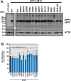
DPP4 harboring polymorphic amino acid residues at the binding interface with MERS-CoV S are robustly expressed. (A) Wildtype (WT) and mutant DPP4 were expressed in 293T cells (cells transfected with empty expression vector served as negative control). Whole cell lysates (WCL) were prepared and analyzed for DPP4 expression by SDS-PAGE under non-reducing conditions and WB using a primary antibody targeting the C-terminal cMYC epitope and a peroxidase-conjugated secondary antibody. Further, expression of beta-actin (ACTB) was analyzed as a loading control. Shown are the expression data from a representative experiment. Numbers at the left indicate the molecular weight in kilodalton (kDa). (B) Quantification of total DPP4 expression in WCL. After normalization of DPP4 band intensities with that of the corresponding ACTB bands. DPP4 WT expression was set as 100% and the relative expression of mutant DPP4 was calculated accordingly. Presented are the combined data of three independent experiments with error bars indicating the SEM. No statistical significance for differences in total expression between WT and mutant DPP4 was observed by one-way analysis of variance with Dunnett’s posttest (p > 0.05, not significant [ns]).
Figure 3.
Figure 3.
DPP4 harboring polymorphic amino acid residues at the binding interface with MERS-CoV S are efficiently transported to the cell surface. (A) Wildtype (WT) and mutant DPP4 were expressed in BHK-21 cells (cells transfected with empty expression vector served as negative control). Surface expressed DPP4 was stained by subsequent incubation of the non-permeabilized cells with a primary antibody that targets the DPP4 ectodomain and an AlexaFluor488-conjugated secondary antibody. Fluorescent signals representing surface-expressed DPP4 were analyzed by flow cytometry and the mean fluorescence intensity (MFI) values for each sample were calculated. For normalization, the MFI value of the negative control was subtracted from all samples. Further, surface expression of DPP4 WT was set as 100% and the relative surface expression of the DPP4 mutants was calculated accordingly. Shown are the combined data of three experiments with error bars indicating the SEM. Statistical significance for differences in surface expression between WT and mutant DPP4 was tested by one-way analysis of variance with Dunnett’s posttest (p > 0.05, not significant; p ≤ 0.05, *). (B) DPP4 surface expression was further analyzed by immunofluorescence analysis. For this, DPP4 WT or DPP4 mutants were expressed in BHK-21 cells grown on coverslips (cells transfected with empty expression vector served as negative control). After fixation of the cells, surface expressed DPP4 was stained by subsequent incubation of non-permeabilized cells with a primary antibody that targets the DPP4 ectodomain and an AlexaFluor568-conjugated secondary antibody. In addition, cellular nuclei were stained with DAPI. Finally, images were taken using a confocal laser scanning microscope at a magnification of 80x.
Figure 4.
Figure 4.
Identification of polymorphic amino acid residues in DPP4 that do not support efficient MERS-CoV S-driven host cell entry. (A) To investigate whether mutant DPP4 support host cell entry driven by MERS-CoV S, we produced vesicular stomatitis virus pseudotype particles (VSVpp) harboring MERS-CoV S (left) or VSV G (control, right). VSVpp were further inoculated on BHK-21 cells expressing wildtype (WT) or mutant DPP4, or cells that were transfected with empty expression vector. At 16 h posttransduction, transduction efficiency was analyzed by measuring the activity of virus-encoded firefly luciferase. Shown are the combined data from three independent experiments (each performed in quadruplicates) for which transduction efficiency of cells expressing DPP4 WT was set as 100%. Error bars indicate the SEM. Statistical significance of differences in transduction efficiency of cells expressing WT or mutant DPP4 was analyzed by one-way analysis of variance with Dunnett’s posttest (p > 0.05, not significant [ns]; p ≤ 0.05, *; p ≤ 0.01, **; p ≤ 0.001, ***).
Figure 5.
Figure 5.
DPP4 harboring polymorphic amino acid residues at the binding interface with MERS-CoV S poorly support replication of live MERS-CoV. Two DPP4 mutants that showed reduced compatibility for MERS-CoV S-driven host cell entry (K267E and A291P) were analyzed in the context of infection and replication of authentic MERS-CoV. For this, BHK-21 cells expressing wildtype (WT) or mutant DPP4, or no DPP4 at all (negative control) were inoculated with MERS-CoV. At 1 h postinfection, the inoculum was removed and the cells were washed before they received fresh culture medium and were further incubated. MERS-CoV replication was analyzed at 0, 24 and 48 h postinfection by determining MERS-CoV genome equivalents (GE) in the culture supernatant (given as GE/ml) by quantitative reverse-transcriptase PCR. Shown are the combined results of three independent experiments (each performed in triplicates). Error bars indicate the SEM. Statistical significance of differences in MERS-CoV replication in cells expressing WT or mutant DPP4 was analyzed by two-way analysis of variance with Dunnett’s posttest (p > 0.05, ns; p ≤ 0.05, *).
Figure 6.
Figure 6.
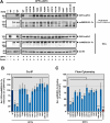
Reduced MERS-CoV S-driven host cell entry is caused by inefficient S protein binding to DPP4 harboring polymorphic amino acid residues. In order to investigate whether reduced MERS-CoV S-driven host cell entry and MERS-CoV replication is due to inefficient MERS-CoV S binding to DPP4 harboring amino acid polymorphisms at the binding interface, we performed co-immunoprecipitation (co-IP) as well as binding experiments with a soluble S protein comprising the S1 subunit of MERS-CoV S fused to the Fc region of human IgG. (A) 293T cells were cotransfected with expression plasmids coding for soluble, Fc-tagged MERS-CoV S1 (solMERS-S1-Fc) and the indicated DPP4 variant containing a C-terminal cMYC-tag. Cells that were transfected only with empty expression vector alone, or empty expression vector instead of either solMERS-S1-Fc or DPP4 served as controls. At 48 h posttransduction, cells were lysed and incubated with protein A sepharose. Next, samples were subjected to SDS-PAGE and Western blot analysis. DPP4 levels were detected via antibodies specific for the cMYC-tag, whereas solMERS-S1-Fc was detected using a peroxidase-coupled anti-human antibody. Similar results were obtained in three individual experiments. Analysis of whole cell lysates (WCL) for expression of solMERS-S1-Fc, DPP4 and ß-actin confirmed comparable ß-actin levels in each sample and comparable expression levels for solMERS-S1-Fc and DPP4. (B) For quantification of MERS-CoV S/DPP4 interaction we first normalized the DPP4 signals against the respective solMERS-S1-Fc signals. Then, MERS-CoV S/DPP4 interaction was set as 100% for wildtype (WT) DPP4 and the relative interaction efficiency for each DPP4 mutant was calculated accordingly. Presented are the mean data from three independent experiments. Error bars indicate the SEM. Statistical significance of differences in MERS-CoV S/DPP4 interaction between WT and mutant DPP4 was analyzed by one-way analysis of variance with Dunnett’s posttest (p > 0.05, ns; p ≤ 0.05, *; p ≤ 0.001, ***). (C) Soluble MERS-CoV S1-Fc was incubated with BHK-21 cells expressing wildtype (WT) or mutant DPP4, or cells transfected with empty expression vector or an ACE2-expression plasmid (controls). To detect bound S protein, the cells were subsequently incubated with an AlexaFluor488-conjugated anti-human antibody directed against the Fc-tag. Fluorescent signals representing bound solMERS-S1-Fc were analyzed by flow cytometry and MFI values for each sample were calculated. For normalization, the MFI value of the negative control (empty expression vector) was subtracted from all samples. Further, binding of solMERS-S1-Fc to cells expressing DPP4 WT was set as 100% and the relative binding to cells expressing the DPP4 mutants was calculated accordingly. Shown are the combined data of five independent experiments with error bars indicating the SEM. Statistical significance of differences in solMERS-S1-Fc binding to cells expressing WT or mutant DPP4 was analyzed by one-way analysis of variance with Dunnett’s posttest (p > 0.05, ns; p ≤ 0.05, *; p ≤ 0.01, **; p ≤ 0.001, ***).

References

    1. Zaki AM, van Boheemen S, Bestebroer TM, et al. . Isolation of a novel coronavirus from a man with pneumonia in Saudi Arabia. N Engl J Med. 2012 Nov 8;367(19):1814–1820. doi: 10.1056/NEJMoa1211721 - DOI - PubMed
    1. WHO . MERS situation update http://www.emro.who.int/health-topics/mers-cov/mers-outbreaks.html2019 [updated 2019–05]. Available from: https://www.who.int/emergencies/mers-cov/en/.
    1. Adney DR, van Doremalen N, Brown VR, et al. . Replication and shedding of MERS-CoV in upper respiratory tract of inoculated dromedary camels. Emerg Infect Dis. 2014 Dec;20(12):1999–2005. doi: 10.3201/eid2012.141280 - DOI - PMC - PubMed
    1. Azhar EI, El-Kafrawy SA, Farraj SA, et al. . Evidence for camel-to-human transmission of MERS coronavirus. N Engl J Med. 2014 Jun 26;370(26):2499–2505. doi: 10.1056/NEJMoa1401505 - DOI - PubMed
    1. Haagmans BL, Al Dhahiry SH, Reusken CB, et al. . Middle East respiratory syndrome coronavirus in dromedary camels: an outbreak investigation. Lancet Infect Dis. 2014 Feb;14(2):140–145. doi: 10.1016/S1473-3099(13)70690-X - DOI - PMC - PubMed

MeSH terms