

Traction force microscopy for understanding cellular mechanotransduction
- PMID: 31964473
- PMCID: PMC7061206
- DOI: 10.5483/BMBRep.2020.53.2.308
Traction force microscopy for understanding cellular mechanotransduction
Abstract
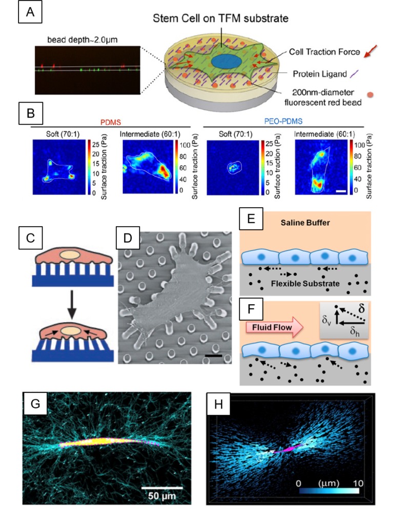
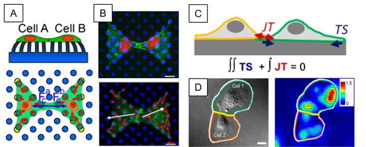
Under physiological and pathological conditions, mechanical forces generated from cells themselves or transmitted from extracellular matrix (ECM) through focal adhesions (FAs) and adherens junctions (AJs) are known to play a significant role in regulating various cell behaviors. Substantial progresses have been made in the field of mechanobiology towards novel methods to understand how cells are able to sense and adapt to these mechanical forces over the years. To address these issues, this review will discuss recent advancements of traction force microscopy (TFM), intracellular force microscopy (IFM), and monolayer stress microscopy (MSM) to measure multiple aspects of cellular forces exerted by cells at cell-ECM and cell-cell junctional intracellular interfaces. We will also highlight how these methods can elucidate the roles of mechanical forces at interfaces of cell-cell/cell-ECM in regulating various cellular functions. [BMB Reports 2020; 53(2): 74-81].
Conflict of interest statement
The authors have no conflicting interests.
Figures
References
Publication types
MeSH terms
Substances
LinkOut - more resources
Full Text Sources
Research Materials
Miscellaneous
