Review
. 2021 Feb 1;11(2):a039362.
doi: 10.1101/cshperspect.a039362.

Opioid-Induced Molecular and Cellular Plasticity of Ventral Tegmental Area Dopamine Neurons

Affiliations
Review

Opioid-Induced Molecular and Cellular Plasticity of Ventral Tegmental Area Dopamine Neurons

Marie A Doyle et al. Cold Spring Harb Perspect Med. .

Abstract

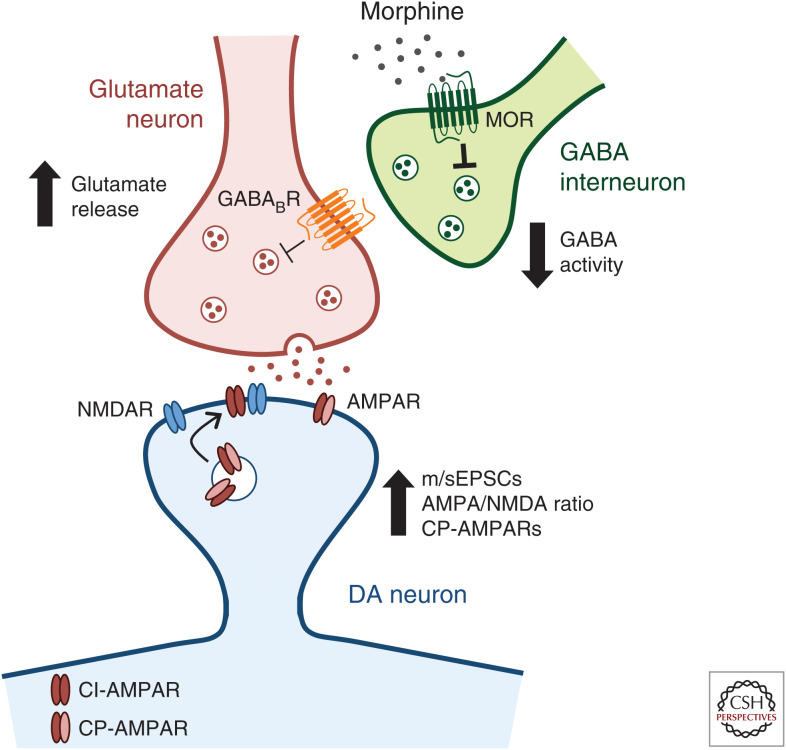
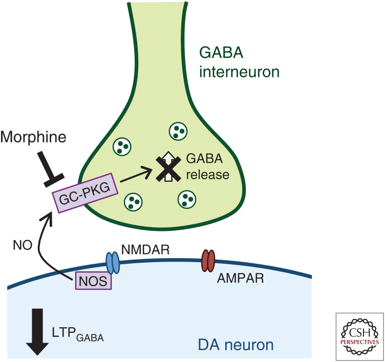
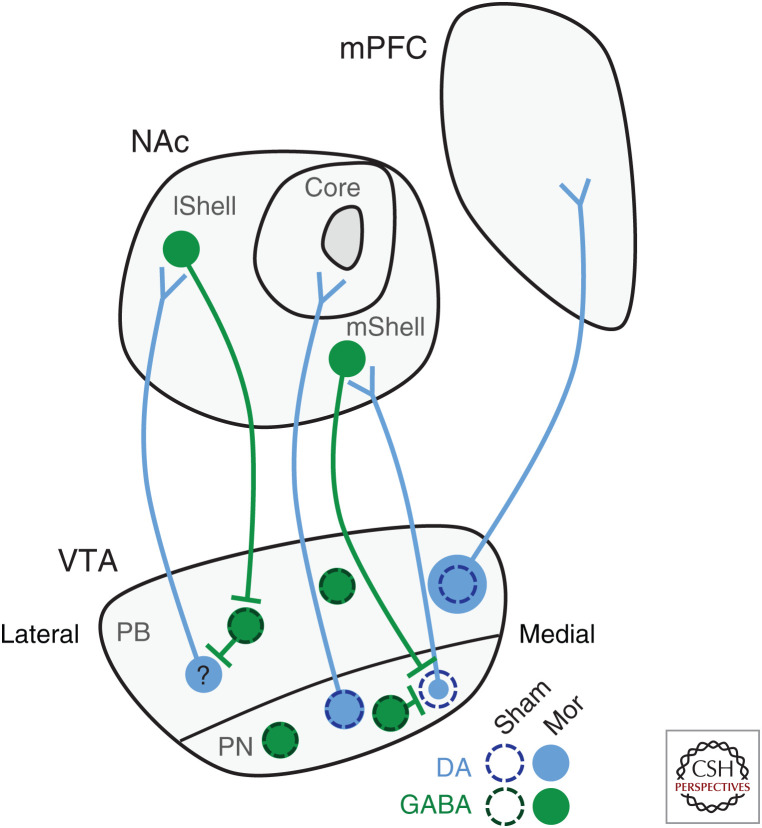
Opioid drugs are highly valued as potent analgesics; however, there are significant risks associated with long-term use because of their abuse liability. Opioids cause changes in ventral tegmental area (VTA) gene expression and cell activity that have been linked to addiction-related behaviors in rodent models. Here, we focus on VTA dopamine (DA) neurons and review the cellular, structural, and synaptic plasticity changes induced by acute and chronic opioid exposure. We also discuss many avenues for future research including determination of whether opioid neuroadaptations are specific for subpopulations of VTA DA neurons. A better understanding of the molecular adaptations within the cells and circuits that drive opioid abuse is crucial for the development of better treatments for substance use disorders and to create novel, safer pain-relieving therapeutics.


Figures

Figure 1.
Figure 1.
Ventral tegmental area (VTA) dopamine (DA)-glutamatergic synaptic plasticity is regulated by acute morphine. Morphine promotes the release of presynaptic glutamate in part through inhibition of VTA γ-aminobutyric acid (GABA) interneurons. Morphine binds to Gi/o-coupled µ-opioid receptors (MORs), causing a hyperpolarization of GABA neurons and decreasing their firing. This decreased GABAergic output onto glutamate neurons, via GABAB receptor signaling, results in increased frequency of miniature and spontaneous excitatory postsynaptic currents (m/sEPSCs) of DA neurons (Baimel and Borgland 2015; Chen et al. 2015). At a second level of DA-glutamatergic plasticity, morphine increases α-amino-3-hydroxy-5-methyl-4-isoxazolepropionic acid receptor/N-methyl-d-aspartic acid receptor (AMPA/NMDA) ratio, consistent with an increase in long-term potentiation (LTP), and causes the exchange of calcium-impermeable GluA2-containing AMPARs (CI-AMPARs) for calcium-permeable GluA2-lacking AMPARs (CP-AMPARs) (Saal et al. 2003; Baimel and Borgland 2015; Authement et al. 2016). Together, these mechanisms of synaptic plasticity increase glutamatergic signaling in the VTA.
Figure 2.
Figure 2.
Acute morphine disrupts ventral tegmental area (VTA) dopamine (DA)-γ-aminobutyric acid (GABA) synaptic plasticity. Acute morphine treatment prevents long-term potentiation of GABAergic (LTPGABA) synapses on VTA DA neurons. LTPGABA derives from NMDAR-dependent release of nitric oxide (NO) via activation of NO synthase (NOS) (Nugent et al. 2007). NO acts as a retrograde messenger to activate guanylate cyclase (GC) in presynaptic GABAergic terminals, resulting in increased GABA release and initiating LTPGABA (Nugent et al. 2007). Morphine prevents LTPGABA and disrupts normal inhibitory signaling in the VTA by blocking NO−GC−protein kinase G (PKG) signaling (Nugent et al. 2007, 2009; Niehaus et al. 2010).
Figure 3.
Figure 3.
Morphine alters ventral tegmental area (VTA) dopamine (DA) soma size in a projection-specific manner. Chronic opioid exposure alters VTA DA, but not γ-aminobutyric acid (GABA), neuron soma size (Sklair-Tavron et al. 1996; Spiga et al. 2003; Chu et al. 2007; Russo et al. 2007; Mazei-Robison et al. 2011). However, the direction of change is projection specific. DA neurons projecting to the nucleus accumbens (NAc) medial shell (mShell) show the decrease in soma size while those projecting to the core are not different from sham controls (Simmons et al. 2019). It remains unknown whether neurons projecting to the NAc lateral shell (lShell) show a decrease in soma size as well. In contrast, VTA DA neurons projecting to the medial prefrontal cortex (mPFC) show a surprising increase in soma size (Simmons et al. 2019), (PB) Parabrachial, (PN) paranigral. (Circuit mapping summarized from Lammel et al. 2014 and Yang et al. 2018.)

References

    1. Authement ME, Langlois LD, Kassis H, Gouty S, Dacher M, Shepard RD, Cox BM, Nugent FS. 2016. Morphine-induced synaptic plasticity in the VTA is reversed by HDAC inhibition. J Neurophysiol 116: 1093–1103. 10.1152/jn.00238.2016 - DOI - PMC - PubMed
    1. Avey D, Sankararaman S, Yim AKY, Barve R, Milbrandt J, Mitra RD. 2018. Single-cell RNA-Seq uncovers a robust transcriptional response to morphine by glia. Cell Rep 24: 3619–3629.e4. 10.1016/j.celrep.2018.08.080 - DOI - PMC - PubMed
    1. Baimel C, Borgland SL. 2015. Orexin signaling in the VTA gates morphine-induced synaptic plasticity. J Neurosci 35: 7295–7303. 10.1523/jneurosci.4385-14.2015 - DOI - PMC - PubMed
    1. Ballantyne JC, LaForge KS. 2007. Opioid dependence and addiction during opioid treatment of chronic pain. Pain 129: 235–255. 10.1016/j.pain.2007.03.028 - DOI - PubMed
    1. Bals-Kubik R, Ableitner A, Herz A, Shippenberg TS. 1993. Neuroanatomical sites mediating the motivational effects of opioids as mapped by the conditioned place preference paradigm in rats. J Pharmacol Exp Ther 264: 489–495. - PubMed

MeSH terms

Substances

LinkOut - more resources