. 2020 Mar 6;295(10):3316-3329.
doi: 10.1074/jbc.RA119.010004. Epub 2020 Jan 21.

A tripartite cooperative mechanism confers resistance of the protein kinase A catalytic subunit to dephosphorylation

Affiliations

A tripartite cooperative mechanism confers resistance of the protein kinase A catalytic subunit to dephosphorylation

Tung O Chan et al. J Biol Chem. .

Abstract

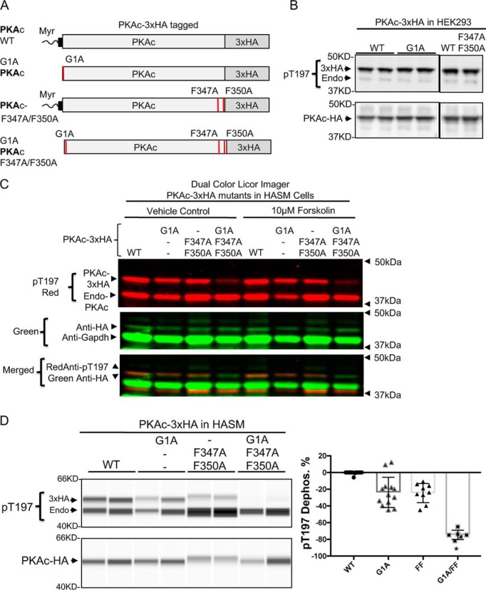
Phosphorylation of specific residues in the activation loops of AGC kinase group (protein kinase A, G, and C families) is required for activity of most of these kinases, including the catalytic subunit of PKA (PKAc). Although many phosphorylated AGC kinases are sensitive to phosphatase-mediated dephosphorylation, the PKAc activation loop uniquely resists dephosphorylation, rendering it "constitutively" phosphorylated in cells. Previous biophysical experiments and structural modeling have suggested that the N-terminal myristoylation signal and the C-terminal FXXF motif in PKAc regulate its thermal stability and catalysis. Here, using site-directed mutagenesis, molecular modeling, and in cell-free and cell-based systems, we demonstrate that substitutions of either the PKAc myristoylation signal or the FXXF motif only modestly reduce phosphorylation and fail to affect PKAc function in cells. However, we observed that these two sites cooperate with an N-terminal FXXW motif to cooperatively establish phosphatase resistance of PKAc while not affecting kinase-dependent phosphorylation of the activation loop. We noted that this tripartite cooperative mechanism of phosphatase resistance is functionally relevant, as demonstrated by changes in morphology, adhesion, and migration of human airway smooth muscle cells transfected with PKAc variants containing amino acid substitutions in these three sites. These findings establish that three allosteric sites located at the PKAc N and C termini coordinately regulate the phosphatase sensitivity of this enzyme. This cooperative mechanism of phosphatase resistance of AGC kinase opens new perspectives toward therapeutic manipulation of kinase signaling in disease.

Keywords: Akt PKB; PKA; cAMP-dependent protein kinase; dephosphorylation; phosphatase; protein kinase B; serine/threonine protein kinase; signal transduction; structure–function.


Conflict of interest statement

The authors declare that they have no conflicts of interest with the contents of this article. The content is solely the responsibility of the authors and does not necessarily represent the official views of the National Institutes of Health

Figures

Figure 1.
Figure 1.
Structural basis for differential phosphatase resistance between PKAc and Akt. A, recombinant (Recomb) PKAc is more resistant to dephosphorylation than recombinant phospho-Akt1 in a cell-free assay. Recombinant PKAc (50 ng) and recombinant Akt1 (50 ng) were incubated with recombinant bacteriophage λ phosphatase (40 units) for 60 min at 30 °C. Images show Akt1 Thr308 and PKAc Thr197 phosphorylation and total Akt and PKAc. Graphs show the percentage of pThr197 dephosphorylation. n = 4/group. *, p < 0.01 for phosphatase versus phosphatase + ATP pocket inhibitor (1 μm A443654). B, amino acid homology comparison between human Akt2 and the human PKA catalytic subunit. Shown is the myristoylated (Myr) PKA–Mn-ATP–cAMP-dependent protein kinase inhibitor peptide structure (PDB code 4DG0). The PKAc myristoylated lipid (light blue), the FXXW motif in the N terminus (N-term, blue), and the C terminus (C-term) FXXF motif (red) form stable and high-affinity contacts.
Figure 2.
Figure 2.
The myristoylated signal and FXXF motif coordinate to protect activation loop phosphorylation in the PKA catalytic subunit. A, to distinguish heterologously expressed PKAc from endogenous PKAc, three copies of the HA epitope were fused with the PKA catalytic subunit at the C terminus (PKAc-3×HA), and mutations were introduced to disrupt the myristoylation signal (G1A) and the c-terminal hydrophobic motif (F347A/F350A). B, transfected HEK293 cells expressing the constructs were propagated for another 4 h in exogenous growth factor–free medium. Protein extracts were subjected to immunoblot analysis using antibodies to phospho-PKA-Thr197 and anti-HA tag antibody. C, PKAc pThr197 and anti-HA detection using the dual-color LI-COR IR image detection system. After 24 h of transfection with the indicated constructs, transfected primary HASM cells were propagated for another 2 h in exogenous growth factor–free medium and stimulated with 10 μm forskolin for 0.5 h as indicated. Shown is immunoblot analysis using antibodies detecting the phosphorylated PKA activation loop (pThr197, red) and anti-HA and anti-GAPDH multiplex (green) showing phosphorylated endogenous and transfected PKAc and GAPDH. D, after 24 h of transfection with the indicated constructs, transfected primary HASM cells were propagated for another 2 h in exogenous growth factor–free medium as indicated. Protein extracts were subjected to immunoblot analysis using the ProteinSimple Wes digital Western system with antibodies to phospho-PKA-Thr197 and anti-HA tag. The graph shows the percentage of HA-PKAc dephosphorylation (Dephos. %). n = 17, 13, 9, and 6 for the indicated groups. *, p < 0.01 G1A/FF versus G1A or FF.
Figure 3.
Figure 3.
N-terminal FXXW contributes to activation loop phosphorylation protection in concert with the C-Terminal FXXF motif. A, the PKAc N-terminal α-helix (blue, aa 13–40) forms stable contacts with the kinase core amino acids. The PKA catalytic subunit fused with three copies of the HA epitope was deleted (aa 13–34 or 24–34). The constructs were transfected into HEK293 cells. Protein lysates were subjected to immunoblot analysis using antibodies for phospho-PKA Thr197 and the HA tag. B, molecular mapping of amino acids affects PKA phosphorylation in the PKAc N terminus. Phe26 and Trp30 regulate Thr197 phosphorylation. HEK293 cells were transfected with PKAc-3×HA WT or the indicated mutants. Protein lysates were subjected to immunoblot analysis using antibodies for phospho-PKA Thr197 and the HA tag. C, the PKA catalytic subunit fused with three copies of the HA epitope were mutated at the N-terminal hydrophobic motif (F26A/W30A) and/or at the C-terminal hydrophobic motif (F347A/F350A). After 24 h of transfection, transfected primary HASM cells were propagated for another 2 h in exogenous growth factor–free medium. Protein extracts were subjected to the ProteinSimple Wes digital Western system using antibodies to phospho-PKA-Thr197 and the anti-HA tag. The graph shows the percentage of HA-PKAc dephosphorylation (Dephos. %). n = 17, 10, 9, and 8 for the indicated groups. *, p < 0.01 FW/FF versus FW or FF.
Figure 4.
Figure 4.
PKAc mutations reduced phosphatase resistance in cell-free assays. A, to define conditions that could rephosphorylate the PKAc mutant at Thr197, the WT and the PKAc F26A/W30A/F347A/F350A mutant were first cotransfected with the kinase PDK1, which phosphorylates the PKA Thr197 site. Then transfected cells were stimulated for 15 min with the phosphatase inhibitor Per-VO4 (100 μm) in the presence of forskolin (FSK). Immunoblot analysis using antibodies detecting phosphorylated PKA-Thr197 showed that combined treatment of forskolin/Per-VO4/PDK1 phosphorylated the PKAc F26A/W30A/F347A/F350A mutant. B, HEK293 cells expressing WT and PKAc F26A/W30A/F347A/F350A mutant constructs were maximally phosphorylated. Flash-frozen cell extracts were prepared on ice with phosphatase inhibitor–free detergent extraction buffer. The extracts were incubated at 30 °C for 30 min. After incubation, protein extracts were subjected to immunoblot analysis using antibodies detecting phosphorylated PKAc Thr197. The graph shows the percentage of PKAc dephosphorylation at 30 min (Dephos. %). n = 2, 6, 2, and 6 for the indicated groups. *p < 0.05 versus WT 30 min. C, PKAc mutants have enhanced phosphatase sensitivity in vitro. Prephosphorylated WT and the PKAc mutants G1A/F347A/F350A and G1A/F26A/W30A/F347A/F350A were immunoprecipitated and incubated with recombinant phosphatases for 20 min at 30 °C. PKAc expression and pThr197 phosphorylation were analyzed with the ProteinSimple Wes imaging system. The graph shows the percentage of pThr197 dephosphorylation in the immunoprecipitates by phosphatases normalized to total PKA expression. n = 4, 7, 4, 7, 3, and 6 for the indicated groups. *, p < 0.05 versus phosphatase-treated WT.
Figure 5.
Figure 5.
N-terminal and C-terminal motifs coordinate to protect PKAc function. A, the PKA catalytic subunit fused with three copies of the HA epitope were mutated at the activation loop (T197A). The constructs were transfected into HASM cells. A GFP construct was cotransfected to monitor transfection efficiency and imaged at 24 h. HASM cells are shown in ×100 GFP images. GFP images of these cells are shown with their phase-contrast images in Fig. S7A. B, PKAc-3xHA with mutations of the myristoylation signal (G1A) and/or the C-terminal hydrophobic motif (F347A/F350A) were transfected into HASM cells. A GFP construct was cotransfected to monitor transfection efficiency and imaged at 24 h. GFP images of HASM cells are shown at ×40. The graph shows the percentage of round GFP cells in transfected PKAc constructs. Number of counted cell fields: n = 7, 10, 7, 7, 9, and 4 for the indicated groups. *, p < 0.05 versus WT or G1A or FF. C, cell adhesion and spreading were measured using the xCELLigence real-time cell analysis system (ACEA Biosciences). After 24 h of transfection, PKA mutant–transfected HASM cells were detached by trypsinization and plated onto E-Plate 16 (ACEA Bioscience) at 6000 cells/well. A graph of plate impedance monitored every 1 min for 10 h is shown. D, PKA mutants affected cell migration in HASM cells. After 6 h of transfection, 10,000 HASM cells were plated into silicone insert molds with a 500-μm defined cell-free gap (Ibidi). After 10 h of incubation in the mold, the mold barrier was removed. The gap spaces were imaged 72 h after silicon mold removal. GFP images of the gap spaces are shown (×40). The graph shows the number of GFP cells in the migration gap in transfected PKAc constructs. Number of counted cell fields: n = 7 for each of the indicated groups. *, p < 0.05 versus WT.
Figure 6.
Figure 6.
A, identification of three dephosphorylation protection motifs in the PKA catalytic subunit structure. B, the cytosolic or membrane-bound PKA catalytic subunit is constitutively phosphorylated at the activation loop (Thr197 in PKA catalytic subunit α), in part because of formation of a phosphatase-resistant conformation that shields phosphorylated Thr197 from cellular phosphatases. C, coordinated disabling the N-terminal myristoylation and FXXW and the C-Terminal FXXF hydrophobic motifs cooperatively reduces phosphatase resistance of the PKA catalytic subunit.

References

    1. Taylor S. S., Ilouz R., Zhang P., and Kornev A. P. (2012) Assembly of allosteric macromolecular switches: lessons from PKA. Nat. Rev. Mol. Cell Biol. 13, 646–658 10.1038/nrm3432 - DOI - PMC - PubMed
    1. Billington C. K., Ojo O. O., Penn R. B., and Ito S. (2013) cAMP regulation of airway smooth muscle function. Pulm. Pharmacol. Ther. 26, 112–120 10.1016/j.pupt.2012.05.007 - DOI - PMC - PubMed
    1. Chan T. O., Pascal J. M., Armen R. S., and Rodeck U. (2012) Autoregulation of kinase dephosphorylation by ATP binding in AGC protein kinases. Cell Cycle 11, 475–478 10.4161/cc.11.3.19059 - DOI - PMC - PubMed
    1. Huse M., and Kuriyan J. (2002) The conformational plasticity of protein kinases. Cell 109, 275–282 10.1016/S0092-8674(02)00741-9 - DOI - PubMed
    1. Pearce L. R., Komander D., and Alessi D. R. (2010) The nuts and bolts of AGC protein kinases. Nat. Rev. Mol. Cell Biol. 11, 9–22 10.1038/nrm2822 - DOI - PubMed

Publication types

MeSH terms

Substances

LinkOut - more resources