. 2020 Jan 21;11(1):e02956-19.
doi: 10.1128/mBio.02956-19.

Inhibition of HIV Replication by Apolipoprotein A-I Binding Protein Targeting the Lipid Rafts

Affiliations

Inhibition of HIV Replication by Apolipoprotein A-I Binding Protein Targeting the Lipid Rafts

Larisa Dubrovsky et al. mBio. .

Erratum in

Abstract

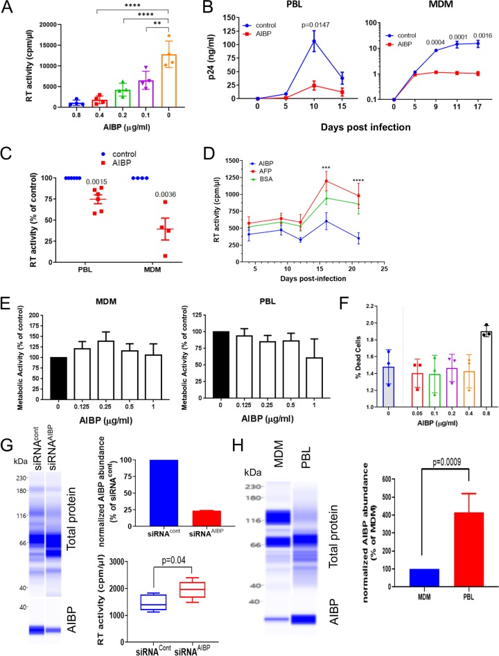
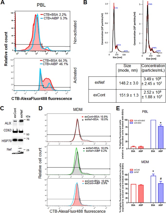
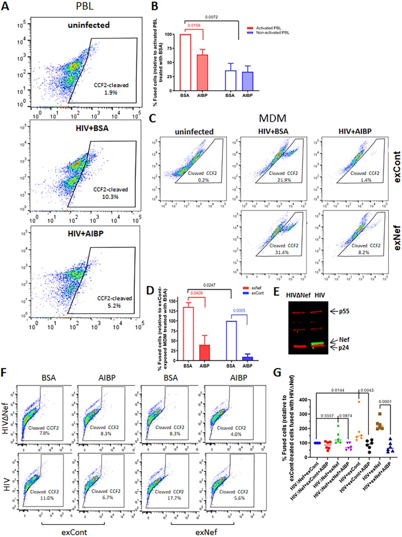
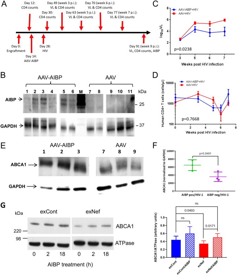
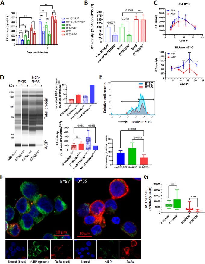
Apolipoprotein A-I binding protein (AIBP) is a protein involved in regulation of lipid rafts and cholesterol efflux. AIBP has been suggested to function as a protective factor under several sets of pathological conditions associated with increased abundance of lipid rafts, such as atherosclerosis and acute lung injury. Here, we show that exogenously added AIBP reduced the abundance of lipid rafts and inhibited HIV replication in vitro as well as in HIV-infected humanized mice, whereas knockdown of endogenous AIBP increased HIV replication. Endogenous AIBP was much more abundant in activated T cells than in monocyte-derived macrophages (MDMs), and exogenous AIBP was much less effective in T cells than in MDMs. AIBP inhibited virus-cell fusion, specifically targeting cells with lipid rafts mobilized by cell activation or Nef-containing exosomes. MDM-HIV fusion was sensitive to AIBP only in the presence of Nef provided by the virus or exosomes. Peripheral blood mononuclear cells from donors with the HLA-B*35 genotype, associated with rapid progression of HIV disease, bound less AIBP than cells from donors with other HLA genotypes and were not protected by AIBP from rapid HIV-1 replication. These results provide the first evidence for the role of Nef exosomes in regulating HIV-cell fusion by modifying lipid rafts and suggest that AIBP is an innate factor that restricts HIV replication by targeting lipid rafts.IMPORTANCE Apolipoprotein A-I binding protein (AIBP) is a recently identified innate anti-inflammatory factor. Here, we show that AIBP inhibited HIV replication by targeting lipid rafts and reducing virus-cell fusion. Importantly, AIBP selectively reduced levels of rafts on cells stimulated by an inflammatory stimulus or treated with extracellular vesicles containing HIV-1 protein Nef without affecting rafts on nonactivated cells. Accordingly, fusion of monocyte-derived macrophages with HIV was sensitive to AIBP only in the presence of Nef. Silencing of endogenous AIBP significantly upregulated HIV-1 replication. Interestingly, HIV-1 replication in cells from donors with the HLA-B*35 genotype, associated with rapid progression of HIV disease, was not inhibited by AIBP. These results suggest that AIBP is an innate anti-HIV factor that targets virus-cell fusion.

Keywords: AIBP; HIV; HLA; Nef; exosomes; extracellular vesicles; fusion; lipid rafts.


Figures

FIG 1
FIG 1
AIBP inhibits HIV-1 replication. (A) Quadruplicate wells of monocyte-derived macrophages (MDMs) were infected with HIV-1ADA, and virus production was measured on day 12 postinfection by analysis of RT activity in culture supernatant. Bars show means ± standard deviations (SD). Statistical analysis was done by Dunnett’s multiple-comparison test. **, P = 0.0013; ****, P < 0.0001. (B) PHA-activated PBLs or MDMs from one representative donor were infected in triplicate wells with HIV-1LAI or HIV-1ADA strains, respectively, cultured in the presence or absence of recombinant AIBP (0.2 μg/ml), and HIV replication was followed by measuring p24 levels in culture supernatants. Holm-Sidak-adjusted P values from multiple-comparison test are shown. (C) The experiment was performed as described for panel B with PHA-activated PBLs from 6 donors and MDMs from 4 donors. Virus replication was followed by analysis of RT activity. Results are presented for each donor at the peak of infection as percent RT activity in AIBP-negative (control) culture. Holm-Sidak-adjusted P values are shown. (D) PHA-activated PBLs were infected with T/F strain pCH185.c/K3016 and cultured in the presence or absence of 0.2 μg/ml recombinant AIBP (recombinant AFP was used as a control). Virus replication was followed by analysis of RT activity. Results show means ± SD (n = 4). Holm-Sidak-adjusted P value is shown. (E) PHA-activated PBLs and MDMs were exposed to the indicated concentrations of recombinant AIBP for 3 days, the cytotoxic effect of AIBP was measured by MTT assay, and the results are presented as percent metabolic activity of AIBP-negative (control) cultures. Bars show means ± SD (n = 4). (F) PHA-activated PBLs were treated with AIBP as described for panel C, and percentages of live cells were measured by flow cytometry using a LIVE/DEAD Fixable Aqua kit (Invitrogen). Bars show means ± SD (n = 3). (G) PHA-activated PBLs were treated with AIBP-targeting or control Accell siRNA for 72 h. AIBP abundance was measured by ProteinSimple Western blotting (left panel) and normalized against total protein, and data are presented relative to the results seen with cells treated with siRNACont (right top panel). Cells were infected in 5 wells with HIV-1LAI and cultured for 3 days, and HIV production was measured by analysis of RT activity in culture supernatant (right bottom panel). P values were calculated by unpaired t test (n = 5). (H) PBLs and MDMs from the same donor were analyzed in triplicate for AIBP by ProteinSimple Western blotting (left panel). Relative abundances of AIBP normalized against total protein are shown in the right panel. P values were calculated by unpaired t test (n = 3).
FIG 2
FIG 2
AIBP regulates abundance of lipid rafts. (A) PBLs from a representative donor were stimulated with PHA or left unstimulated, stained with fluorescently labeled cholera toxin subunit B, and analyzed by flow cytometry. (B) Representative analysis of vesicle size and concentration in exosome samples from supernatants of HEK293 T cells transfected with Nef (exNef) or empty vector (exCont) by Nanosight (top panels). Means ± standard errors of the means (SEM) of vesicle size (in nanometers) and vesicle concentration (in particles per milliliter) are shown in the bottom panel. (C) Vesicles were analyzed by Western blotting for the exosomal marker Alix, tetraspanin CD63, cytosolic marker HSP70, and Nef. (D) MDMs from a representative donor were treated with exNef or exCont in the presence of AIBP or BSA, and lipid rafts were analyzed as described for panel A. (E) Lipid rafts were analyzed as described for panels A and D. Results are presented for experiments performed with cells from 4 different donors. (Left panel) *, P = 0.0088 (unpaired t test performed with Holm-Sidak adjustment, relative to activated PBLs treated with BSA). (Right panel) *, P = 0.0038 (relative to cells treated with exCont and BSA); #, P = 0.0129 (relative to MDMs treated with exNef and BSA; ordinary one-way ANOVA with Tukey’s adjustment for multiple comparisons).
FIG 3
FIG 3
AIBP inhibits HIV fusion with target cells. (A) PBLs were activated with PHA for 48 h, treated with 0.2 μg/ml recombinant AIBP (or BSA as a control) for another 48 h, and exposed to BlaM-Vpr carrying HIV-1NL4-3 in the presence or absence of recombinant AIBP. Percentages of fused cells (cleaved CCF-2) were determined by flow cytometry. (B) Results of fusion analysis of PBLs from 4 donors (means ± SD) are presented relative to fusion of activated PBLs treated with BSA, taken as 100%. P values were calculated by multiple t test with Bonferroni-Dunn adjustment (BSA versus AIBP groups) or one-way ANOVA with Tukey’s adjustment (comparison of individual treatments); only significant values are shown. (C) MDMs were exposed to control exosomes (exCont) or Nef exosomes (exNef) for 48 h in the presence of 0.2 μg/ml recombinant AIBP (or BSA as a control) and were then infected with BlaM-Vpr carrying HIV-1NL(AD8) in the presence or absence of recombinant AIBP. Percentages of fused cells were determined as described for panel A. (D) Results of fusion analysis of MDMs from 3 donors (means ± SD) are presented relative to fusion of exCont-exposed cells treated with BSA, taken as 100%. P values were calculated by multiple t test with Bonferroni-Dunn adjustment for multiple comparisons. (E) Western blot for Nef (green) and p55 (red) of HEK293T cells transfected with vectors expressing Nef-positive and Nef-negative HIV-1. (F) MDMs were exposed to exCont or exNef as described for panel C and infected with BlaM-Vpr carrying Nef-positive or Nef-deficient HIV-1. Fusion was analyzed as described for panel A. (G) An experiment was performed as described for panel F with MDMs from 6 donors. Results are presented for each donor relative to fusion of exCont-treated MDM with HIVΔNef. P values were calculated by repeated-measures ANOVA with Bonferroni correction for multiple comparisons.
FIG 4
FIG 4
AIBP reduces HIV load and reverses ABCA1 downregulation in liver cells. (A) Timeline of the in vivo experiment. (B) Western blot analysis of livers from hu-mice infected with AAV and AAV-AIBP. (C) Viral load analysis of hu-mice. *, P = 0.0238 (by a 2-way ANOVA). (D) Analysis of human CD4+ cells in hu-mice. No significant differences were detected by 2-way ANOVA. (E) Western blot for AIBP and GAPDH (glyceraldehyde-3-phosphate dehydrogenase) in livers from hu-mice infected with HIV-1 and AAV-AIBP (mice 1, 2, and 3) and hu-mice infected with HIV-1 and empty AAV (mice 7, 8, and 9). (F) Quantitation of the blot in panel E. P values were calculated by unpaired t test. (G) HepG2 cells were treated for 48 h with exCont or exNef and were then incubated for 18 h in the presence or absence of AIBP (0.2 μg/ml). Total ABCA1 and K,Na ATPase (loading control) levels were assayed by Western blotting (left panel), and images from 6 independent experiments were quantified by ImageJ (right panel). Results present means ± SD. Significance was calculated by 1-way ANOVA with Tukey’s multiple-comparison adjustment. ns, not statistically significant.
FIG 5
FIG 5
Anti-HIV effect of AIBP is reduced in cells from HLA-B*35 donors. (A) PHA-activated PBMCs from donors with HLA-B*35, HLA-B*57, and non-B*35,B*57 genotypes were infected with HIV-1LAI and incubated in the presence or absence of recombinant AIBP. Virus replication was followed by analysis of RT activity. Results are presented for donors B*35/55 (B*35), B*51/57 (B*57), and B*27/38 (non-B*35,B*57). Results are presented as means ± SD of results from 5 replicates. *, P = 0.01; **, P < 0.01; ***, P < 0.001 (by multiple-comparison tests with Holm adjustment). (B) The experiment was performed as described for panel A with cells from 3 donors of each genotype. Results (means ± SD) are presented relative to cells from non-B*35,57 donor (taken as 100%) at the time point corresponding to the peak of infection (day 5 p.i.). P values were calculated by ordinary one-way ANOVA with Tukey’s multiple-comparison adjustment. (C) PHA-activated PBMCs from HLA-B*35 or non-B*35 donors were infected with T/F strain pCH185.c/K3016 and cultured in the presence or absence of 0.2 μg/ml recombinant AIBP or BSA. Virus replication was followed by analysis of RT activity. Results show means ± SD (n = 4). Significance was calculated by multiple t tests with Holm-Sidak correction for multiple comparisons. ***, P < 0.001. (D) PHA-activated PBMCs from HLA-B*35 and non-B*35 donors were treated with AIBP-targeting (siRNAAIBP) or control (siRNACont) Accell siRNA. AIBP abundance was measured by ProteinSimple Western blotting (left panel) and normalized against total protein, and results are presented relative to non-B*35 cells treated with siRNACont (right top panel). Cells were infected with HIV-1 LAI, and RT activity in culture supernatant was measured on day 4 postinfection (right bottom panel). Results show means ± SD of 4 replicates. Ordinary one-way ANOVA with Tukey’s adjustment for multiple comparisons was used to calculate P values. (E) (Top panel) Binding of recombinant AIBP to cells with different HLA-B genotypes was analyzed by flow cytometry using anti-His antibody. (Bottom panel) Quantitation of AIBP binding to cells from 4 different donors each of genotypes B*35 and non-B*35,B*57 and 3 donors of genotype B*57. P values were calculated by multiple t tests, with post hoc Holm adjustment for multiple comparisons. (F) Binding of recombinant AIBP to cells with HLA-B*35 and HLA-B*57 genotype was analyzed by fluorescence microscopy using Alexa Fluor 555-conjugated CTB for lipid rafts (red), FITC-conjugated anti-His antibody for AIBP (green), and DAPI for nuclei (blue). (G) Quantification of MFI on 108 cells with HLA-B*35 and 159 cells with HLA-B*57 genotype using Volocity software. ****, P < 0.0001 (calculated by multiple t tests with Holm-Sidak correction for multiple comparisons).

References

    1. Ritter M, Buechler C, Boettcher A, Barlage S, Schmitz-Madry A, Orso E, Bared SM, Schmiedeknecht G, Baehr CH, Fricker G, Schmitz G. 2002. Cloning and characterization of a novel apolipoprotein A-I binding protein, AI-BP, secreted by cells of the kidney proximal tubules in response to HDL or ApoA-I. Genomics 79:693–702. doi:10.1006/geno.2002.6761. - DOI - PubMed
    1. Marbaix AY, Noel G, Detroux AM, Vertommen D, Van Schaftingen E, Linster CL. 2011. Extremely conserved ATP- or ADP-dependent enzymatic system for nicotinamide nucleotide repair. J Biol Chem 286:41246–41252. doi:10.1074/jbc.C111.310847. - DOI - PMC - PubMed
    1. Niehaus TD, Elbadawi-Sidhu M, Huang L, Prunetti L, Gregory JF III, de Crecy-Lagard V, Fiehn O, Hanson AD. 2018. Evidence that the metabolite repair enzyme NAD(P)HX epimerase has a moonlighting function. Biosci Rep 38:BSR20180223. doi:10.1042/BSR20180223. - DOI - PMC - PubMed
    1. Gu Q, Yang X, Lv J, Zhang J, Xia B, Kim JD, Wang R, Xiong F, Meng S, Clements TP, Tandon B, Wagner DS, Diaz MF, Wenzel PL, Miller YI, Traver D, Cooke JP, Li W, Zon LI, Chen K, Bai Y, Fang L. 2019. AIBP-mediated cholesterol efflux instructs hematopoietic stem and progenitor cell fate. Science 363:1085–1088. doi:10.1126/science.aav1749. - DOI - PMC - PubMed
    1. Fang L, Choi SH, Baek JS, Liu C, Almazan F, Ulrich F, Wiesner P, Taleb A, Deer E, Pattison J, Torres-Vazquez J, Li AC, Miller YI. 2013. Control of angiogenesis by AIBP-mediated cholesterol efflux. Nature 498:118–122. doi:10.1038/nature12166. - DOI - PMC - PubMed

Publication types