. 2020 Feb 4;117(5):2432-2440.
doi: 10.1073/pnas.1910208117. Epub 2020 Jan 21.

Structural elements regulating the photochromicity in a cyanobacteriochrome

Affiliations

Structural elements regulating the photochromicity in a cyanobacteriochrome

Xiuling Xu et al. Proc Natl Acad Sci U S A. .

Erratum in

Abstract

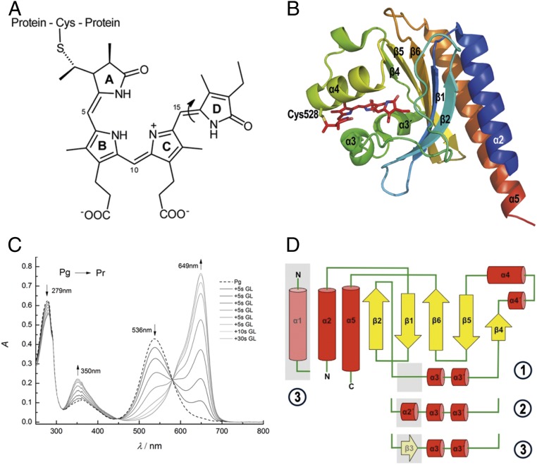
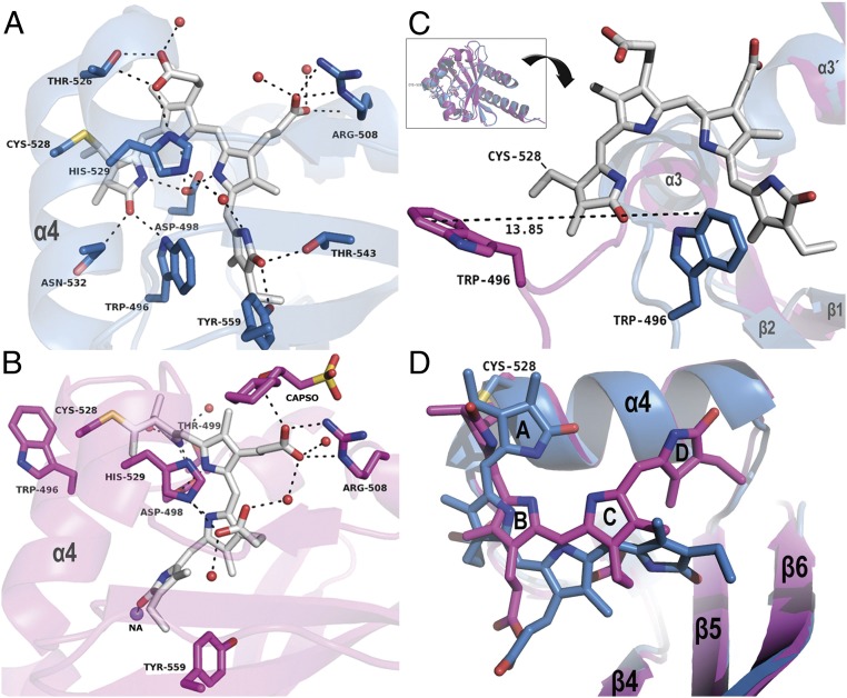
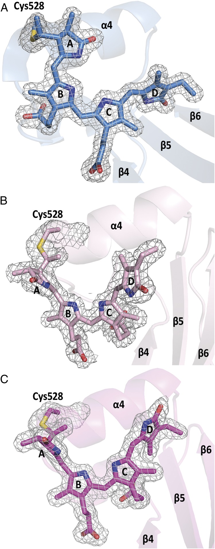
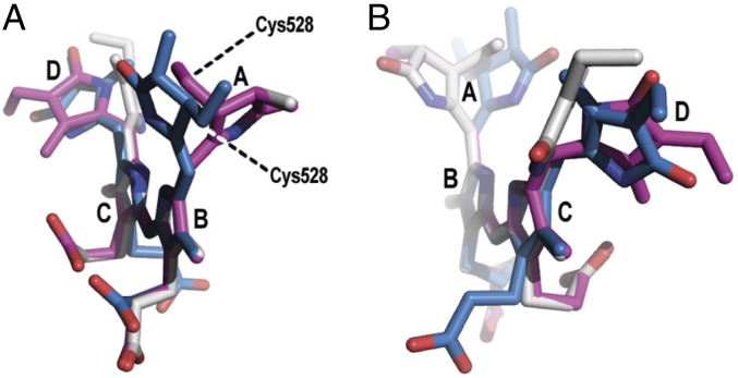
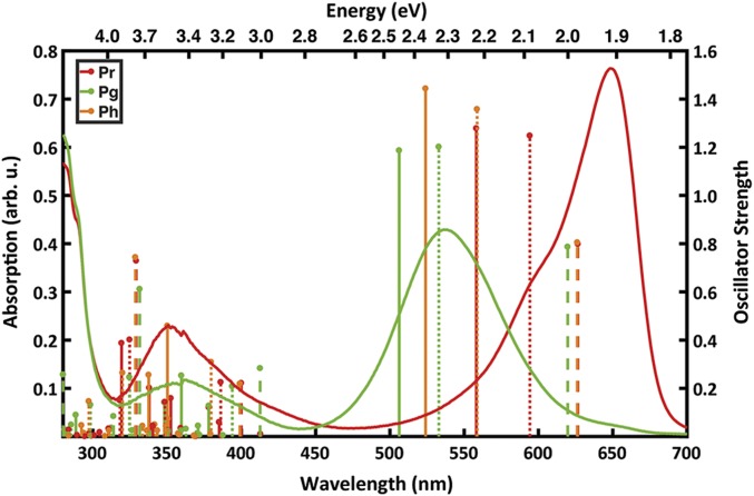
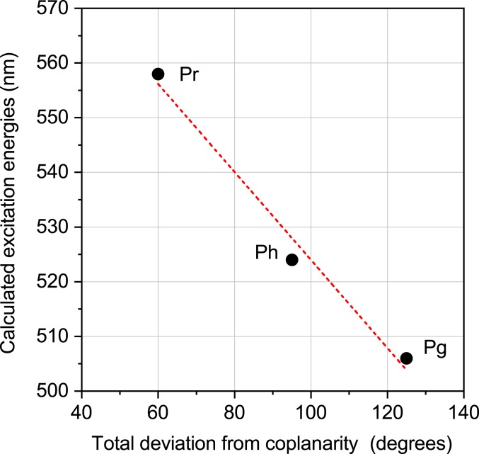
The three-dimensional (3D) crystal structures of the GAF3 domain of cyanobacteriochrome Slr1393 (Synechocystis PCC6803) carrying a phycocyanobilin chromophore could be solved in both 15-Z dark-adapted state, Pr, λmax = 649 nm, and 15-E photoproduct, Pg, λmax = 536 nm (resolution, 1.6 and 1.86 Å, respectively). The structural data allowed identifying the large spectral shift of the Pr-to-Pg conversion as resulting from an out-of-plane rotation of the chromophore's peripheral rings and an outward movement of a short helix formed from a formerly unstructured loop. In addition, a third structure (2.1-Å resolution) starting from the photoproduct crystals allowed identification of elements that regulate the absorption maxima. In this peculiar form, generated during X-ray exposition, protein and chromophore conformation still resemble the photoproduct state, except for the D-ring already in 15-Z configuration and tilted out of plane akin the dark state. Due to its formation from the photoproduct, it might be considered an early conformational change initiating the parental state-recovering photocycle. The high quality and the distinct features of the three forms allowed for applying quantum-chemical calculations in the framework of multiscale modeling to rationalize the absorption maxima changes. A systematic analysis of the PCB chromophore in the presence and absence of the protein environment showed that the direct electrostatic effect is negligible on the spectral tuning. However, the protein forces the outer pyrrole rings of the chromophore to deviate from coplanarity, which is identified as the dominating factor for the color regulation.

Keywords: crystal structure; photochromicity; phytochrome; theoretical chemistry.


Conflict of interest statement

The authors declare no competing interest.

Figures

Fig. 1.
Fig. 1.
Chromophore structure and absorption spectra. (A) Chemical structure of phycocyanobilin chromophore of Slr1393g3. The chromophore is bound covalently to the protein via a thioether bond between Cys528 and the 31 position of PCB. The molecule is shown in the parental-state configuration (Z,Z,Z,s,s,a). The double-bond photoisomerization (double bond between rings C and D) is indicated by an arrow. (B) Cartoon representation of Slr1393g3 structure in its in vitro-assembled parental Pr state (PDB ID code 5DFY). Secondary-structure elements are labeled according to the AnPixJg2 structure (PDB ID code 3W2Z). The PCB chromophore is shown in stick representation; the covalent bond to Cys528 is highlighted. (C) Illumination-induced conversion between the red- (Pr) and green-absorbing (Pg) forms of Slr1393g3. Formation of the red-absorbing form follows stepwise irradiation of Pg (total irradiation time, 80 s; irradiation source, 670-nm LED). (D) Topology of Slr1393g3 in comparison to AnPixJg2: ① Slr1393g3-Pr state, ② Slr1393g3-Pg and the hybrid form, and ③ AnPixJg2 topology. The topology depicts Slr1393g3 in the parental, red-absorbing state. The gray box between β2 and α3 (part of an unstructured loop) converts into a short helical element in the photoproduct and in the photoisomerization hybrid (coined α2′ in the main text). In the parental state of AnPixJg2, this adapts a β-sheet conformation (β3).
Fig. 2.
Fig. 2.
Comparison of the chromophore protein interactions in the Pr and Pg state. (A) Binding site of Slr1393g3-Pr; the chromophore (gray sticks) is in the Z,Z,Z,s,s,a configuration. (B) Binding site of Slr1393g3-Pg; the chromophore (gray sticks) is in the Z,Z,E,s,s,a configuration. In A and B, helix α4 is labeled for clarity; residues interacting with the chromophore are represented as sticks and are labeled individually, water molecules as red spheres, and sodium as purple sphere. Distances are given in SI Appendix, Table S2. (C) The Inset on Top shows an overview of the superimposed structures of Slr1393g3-Pr (blue cartoon; PDB ID code 5DFY) and Slr1393g3-Pg (magenta; PDB ID code 5M82). Below the Inset, the structure is rotated for a detailed view on the movement of Trp496; for clarity, the PCB chromophore in Pg state is omitted; the Pr-Trp is shown in blue, and Pg-Trp in magenta. (D) Detailed view on the chromophore conformation of both states with color coding adapted from C. The PCB molecule in the Pg state is, compared with the Pr state, closer to helix α4 and has moved further into the binding site.
Fig. 3.
Fig. 3.
Chromophore conformations and electron densities in Slr1393g3. (A) Pr state, (B) hybrid state (back isomerized chromophore) showing a highly distorted Z,Z,Z,s,s,a configuration, and (C) Pg photoproduct showing the chromophore in Z,Z,E,s,s,a configuration. Note the changed relative orientation of the PCB molecule in B and C with respect to helix α4 and β-sheets β5, β6. Electron densities are shown for the chromophore and the covalently bound Cys528, contoured at 1σ.
Fig. 4.
Fig. 4.
Superposition of the PCB chromophores in different perspectives. Geometries of PCB in Slr1393g3 in Pr state (blue; PDB ID code 5DFY), hybrid state (gray; PDB ID code 5M85), and Pg state (magenta; PDB ID code 5M82). Ring B and C were used as reference points. A shows the chromophore conformation along rings B–C, (B) along rings C–B. Note the two different orientations of ring A in the Pr state versus the Pg and hybrid states. The D ring is oriented differently in the hybrid state, compared to the Pr and Pg states.
Fig. 5.
Fig. 5.
Experimental absorption spectra and calculated excitation energies of Slr1393g3. The experimental absorption spectra (9) are shown as lines for Pr (red) and Pg (green). The calculated excitation energies are represented as sticks for the Pr (red), Pg (green), and the hybrid Ph (orange) forms. The vacuo models are represented by dashed lines, strained models by dotted lines, and protein models as solid sticks. The height of each stick is proportional to the oscillator strength of the corresponding transition.
Fig. 6.
Fig. 6.
Relationship between the calculated QM/MM excitation energies for three structures and the coplanarity of the chromophore. Coplanarity is defined by summing the absolute values of the dihedrals between rings A/B, B/C, and C/D as defined in ref. .

References

    1. Ikeuchi M., Ishizuka T., Cyanobacteriochromes: A new superfamily of tetrapyrrole-binding photoreceptors in cyanobacteria. Photochem. Photobiol. Sci. 7, 1159–1167 (2008). - PubMed
    1. Rockwell N. C., Lagarias J. C., A brief history of phytochromes. ChemPhysChem 11, 1172–1180 (2010). - PMC - PubMed
    1. Anders K., Essen L. O., The family of phytochrome-like photoreceptors: Diverse, complex and multi-colored, but very useful. Curr. Opin. Struct. Biol. 35, 7–16 (2015). - PubMed
    1. Montgomery B. L., Lagarias J. C., Phytochrome ancestry: Sensors of bilins and light. Trends Plant Sci. 7, 357–366 (2002). - PubMed
    1. Müller M. G., Lindner I., Martin I., Gärtner W., Holzwarth A. R., Femtosecond kinetics of photoconversion of the higher plant photoreceptor phytochrome carrying native and modified chromophores. Biophys. J. 94, 4370–4382 (2008). - PMC - PubMed

Publication types