

Eosinophilic cytoplasmic inclusions in type 2 papillary renal cell carcinoma
- PMID: 31965115
- PMCID: PMC8145672
- DOI: 10.32074/1591-951X-28-19
Eosinophilic cytoplasmic inclusions in type 2 papillary renal cell carcinoma
Abstract
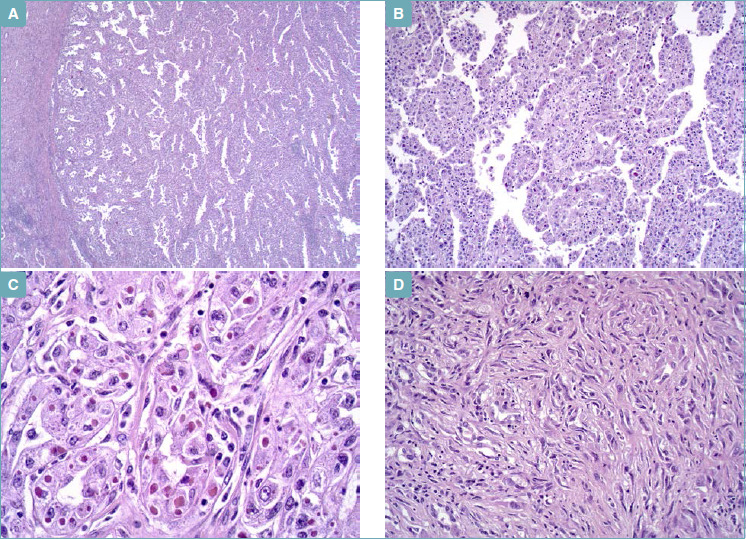
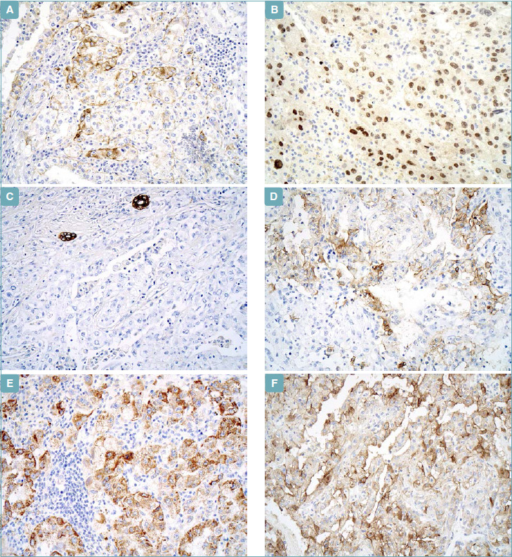
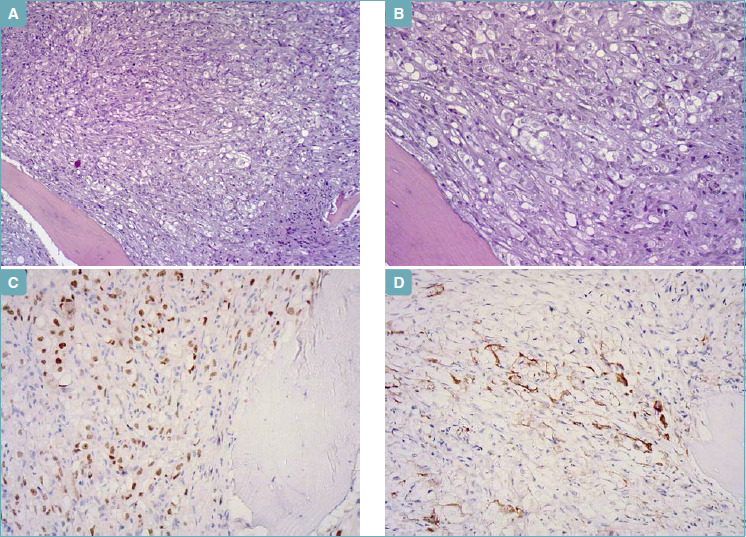
A case of a patient with type 2 papillary renal cell carcinoma with eosinophilic cytoplasmatic inclusions is presented. About 50% of tumor cells were characterized by a well-circumscribed intra-cytoplasmatic round-to-oval or irregular inclusion/globule. Inclusions were 7-30 micron in diameter. They were glassy and pale to slightly eosinophilic in color in hematoxylin and eosin, were stained red by trichrome and were negative for periodic acid-Schiff reaction. Immunohistochemically, globules were negative for PAX8, epithelial membrane antigen, Carbonic Anhydrase IX, pan-cytokeratin (AE1/AE3), CD10, S100 protein, α-smooth-muscle actin, cytokeratin 7 and cytokeratin 34βE12. Glassy hyaline globules were not detected in any adjacent normal kidney cells. The presence of eosinophilic cytoplasmic inclusions in renal cell carcinoma, especially in papillary renal cell carcinoma, has been rarely emphasized in the literature. In this article, we review similar cases in the literature and discuss the nature of eosinophilic globules.
Keywords: Aggressive behavior; Eosinophilic cytoplasmatic inclusions; Pppillary renal cell carcinoma; Sarcomatoid differentiation.
Copyright © 2019 Società Italiana di Anatomia Patologica e Citopatologia Diagnostica, Divisione Italiana della International Academy of Pathology.
Conflict of interest statement
None declared.
Figures
References
-
- Delahunt B, Algaba F, Eble J, et al. . Papillary renal cell carcinoma. Moch H, Humphrey PA, Ulbright TM, et al., eds. WHO Classification of Tumours Of the Urinary System nd Male Genital Organs. 4th ed. Lyon, France; IARC Press; 2016.
-
- Murphy WM, Grignon DJ, Perlman EJ. Tumors of the kidney, bladder, and related urinary structures. AFIP - Atlas of Tumor Pathology. 4th Series; Fascicle 1. Washington DC: 2004.
-
- Renshaw AA, Zhang H, Corless CL, Fletcher JA, Pinis MR. Solid variants of papillary (chromophil) renal cell carcinoma: clinicopathologic and genetic features. Am J Surg Pathol 1997;21:1203-9. https://doi.org/10.1097/00000478-199710000-00011 10.1097/00000478-199710000-00011 - DOI - PubMed
-
- Delahunt B, Eble JN. Papillary renal cell carcinoma: a clinicopathologic and immunohistochemical study of 105 tumors. Mod Pathol 1997;10:537-44. - PubMed
-
- Delahunt B, Eble JN, McCredie MR, et al. . Morphologic typing of papillary renal cell carcinoma: comparison of growth kinetics and patients survival in 66 cases. Hum Pathol 2001;32:590-5. https://doi.org/10.1053/hupa.2001.24984 10.1053/hupa.2001.24984 - DOI - PubMed
Publication types
MeSH terms
LinkOut - more resources
Full Text Sources
Medical
Research Materials
Miscellaneous
