. 2020 Apr 6;48(6):3001-3013.
doi: 10.1093/nar/gkaa022.

Poly(ADP-ribosyl)ation mediates early phase histone eviction at DNA lesions

Affiliations

Poly(ADP-ribosyl)ation mediates early phase histone eviction at DNA lesions

Guang Yang et al. Nucleic Acids Res. .

Abstract

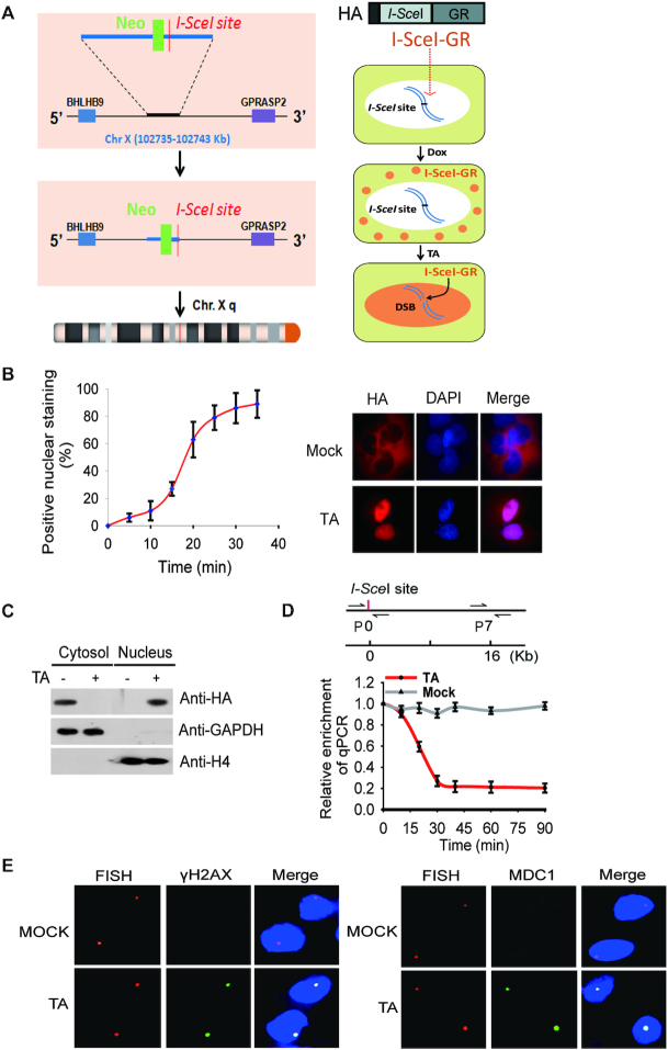
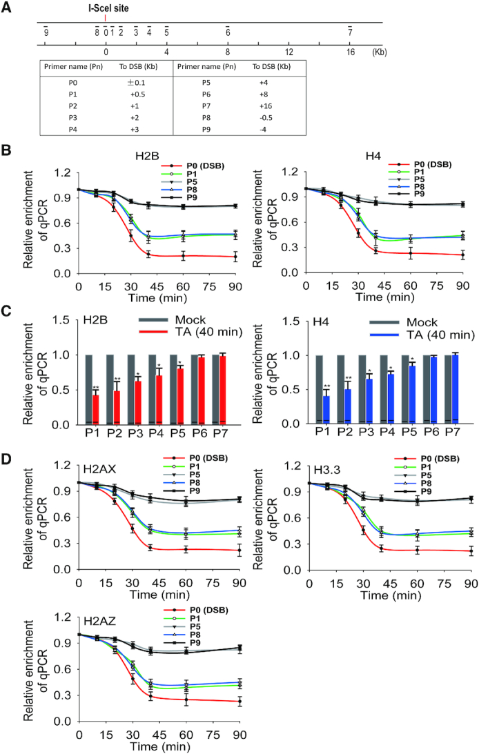
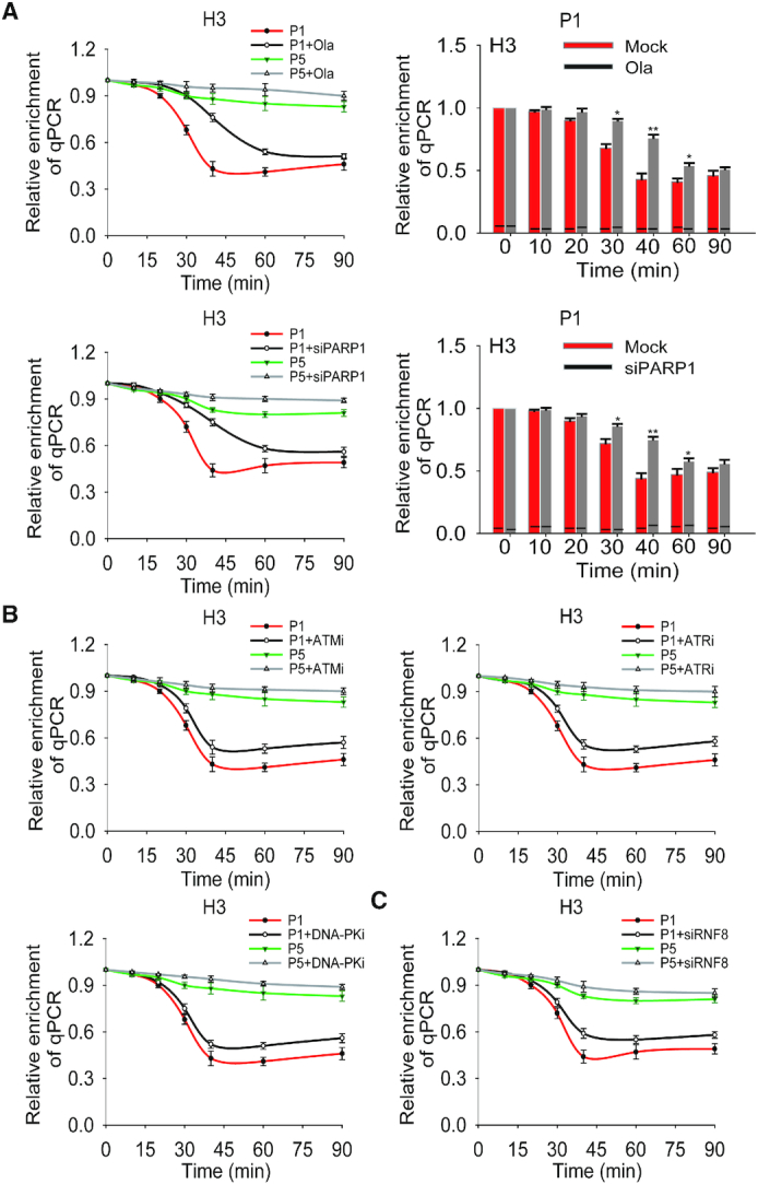
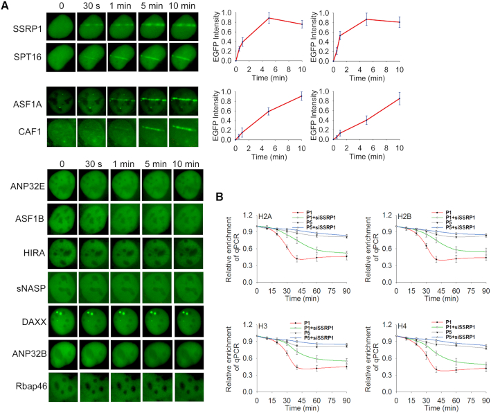
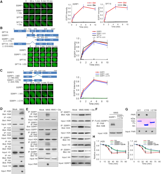
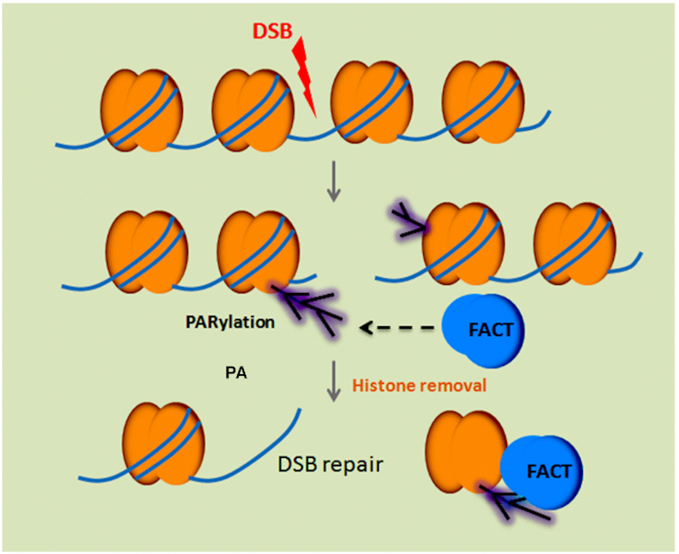
Nucleosomal histones are barriers to the DNA repair process particularly at DNA double-strand breaks (DSBs). However, the molecular mechanism by which these histone barriers are removed from the sites of DNA damage remains elusive. Here, we have generated a single specific inducible DSB in the cells and systematically examined the histone removal process at the DNA lesion. We found that histone removal occurred immediately following DNA damage and could extend up to a range of few kilobases from the lesion. To examine the molecular mechanism underlying DNA damage-induced histone removal, we screened histone modifications and found that histone ADP-ribosylation was associated with histone removal at DNA lesions. PARP inhibitor treatment suppressed the immediate histone eviction at DNA lesions. Moreover, we examined histone chaperones and found that the FACT complex recognized ADP-ribosylated histones and mediated the removal of histones in response to DNA damage. Taken together, our results reveal a pathway that regulates early histone barrier removal at DNA lesions. It may also explain the mechanism by which PARP inhibitor regulates early DNA damage repair.


Figures

Figure 1.
Figure 1.
I-SceI induces a solo DSB in HCT116 cells. (A) A schematic diagram shows that an I-SceI site is inserted into the X chromosome of HCT116 cells using gene targeting approach. I-SceI is fused with a glucocorticoid receptor (GR) and an HA tag, and the expression of the fusion protein is controlled by Doxycycline (Dox). The relocation of the fusion protein from the cytoplasm to the nucleus is dictated by triamcinolone acetonide (TA) treatment. (B) Translocation of I-SceI-GR from cytoplasm to nucleus in response to the TA treatment. The relocation kinetics of I-SceI-GR was measured by immunofluorescence staining staining with anti-HA antibody following TA treatment. The percentage of positive nuclear staining is shown in the histograms. (C) Localization of I-SceI-GR was examined by Western blotting with indicated antibodies. GAPDH and Histone 4 (H4) were used as controls of protein loading for cytosol and nuclear fractions, respectively. (D) I-SceI-GR induced DSB was examined by qPCR. The primers at the ‘0’ position flank both sides of DSB. The primer set #7 at 16 kb downstream of the DSB was used as the reference control. (E) Upon TA induction, I-SceI-GR induces a DSB in the nucleus. DNA probe was used to map the I-SceI site (red dot), which was also stained with anti-γH2AX antibody or anti-MDC1 antibody to indicate the DSB (green dot).
Figure 2.
Figure 2.
Histone eviction at vicinity of I-SceI-induced DSB. (A) A schematic diagram shows the positions of primer sets for ChIP assays with respect to DSB site. The sequences of the primers used in this study were listed in Supplemental Table 1. (B) Removal of nucleosomal histones at the vicinity of I-SceI-induced DSB. The histones H2B and H4 eviction was examined by ChIP assays with anti-H2B or anti-H4 antibodies and qPCR with different sets of primers. The results were analyzed and shown as mean ± SD. (C) Relative enrichment of H2B and H4 are summarized in the histograms. Data were shown as mean ± SD. * P < 0.05, ** P < 0.01. (D) Removal of histones H2AX, H3.3 and H2AZ at the DSB. ChIP assays were carried out with anti-H2AX, anti-H3.3 and anti-H2AZ antibodies for the histone removal analyses. The results were analyzed and shown as mean ± SD.
Figure 3.
Figure 3.
PARP inhibition delays histone removal at the I-SceI-induced DSB. (A) The impact of PARylation in histone removal at the I-SceI-induced DSB. The cells were treated with or without 1 μM olaparib or PARP1 siRNA. The histone removal was examined by ChIP assays with anti-histone H3 antibody. The qPCR assays were performed with primer set #1 and #5 (shown as P1 and P5 in Supplemental Table 1) at indicated time points. The results of qPCR from P1 were summarized in the histogram and shown as mean ± SD (right panel) (*P < 0.05; ** P < 0.01). (B) The effect of PI-3 like kinase-mediated phosphorylation on histone removal at the I-SceI-induced DSB. The histone removal was examined by ChIP assays following cells were treated with or without ATM inhibitor (ATMi, 5 μM KU60019), ATR inhibitor (ATRi, 1 μM VE822) or DNA-PK inhibitor (DNA-PKi, 1 μM NU7441). Results were analyzed and shown as mean ± SD. (C) The impact of ubiquitination on histone removal at the I-SceI-induced DSB. The cells treated with or without RNF8 siRNA were used for assessing histone removal by ChIP assays. Relative enrichment was shown as mean ± SD.
Figure 4.
Figure 4.
Histone chaperones are recruited to DNA lesions. (A) The relocation kinetics of SSRP1, SPT16, ASF1A, and CAF1 to DNA lesions. GFP tagged histone chaperones were expressed in U2OS cells. The relocation was monitored in a time course following laser microirradiation. The relative EGFP intensity is shown as mean ± SD. (B) Loss of the FACT complex impairs histone removal from the DSB. The histone chaperone SSRP1 knockdown cells were harvested following I-SceI-induced DSB and subjected to ChIP assay using anti-H2A, anti-H2B, anti-H3 or anti-H4 antibodies. The samples were examined by qPCR with primer set #1 and #5 to examine histone removal. Relative enrichment of histones at the DSB was shown in the graphs. **P < 0.01.
Figure 5.
Figure 5.
The FACT complex recognizes PARylation at DNA lesions. (A) The FACT complex recognizes PARylation at DNA lesions. U2OS cells expressing GFP-SSRP1 or GFP-SPT16 were pretreated with or without 1 μM olaparib for 2 hours, followed by laser microirradiation. The relocation kinetics of GFP-tagged SSRP1 and SPT16 were monitored in a time course. Results were shown as mean ± SD. (B) SSRP1 mediates the recruitment of the FACT complex to DNA lesions. The DD mutants of SSRP1 and SPT16 with an N-terminal GFP tag were expressed in U2OS cells. The recruitment kinetics was examined. (C) Mapping the domain of SSRP1 that recognizes PARylation. GFP-SSRP1 with deletion of the MD domain (MD) or the C-terminal region (CTR) was expressed in U2OS cells. The recruitment kinetics was examined. (D) Histones are PARylated following DNA damage. 293T cells were treated with or without 1 mM MMS. Damage-induced histone PARylation was examined with indicated antibodies. (E) SSRP1 recognizes PARylated histones. 293T cells were treated with 1 mM MMS in the presence or absence of olaparib (1 μM). SSRP1-associated PARylated histones were analyzed with sequential IP with anti-SSRP1 and anti-PAR antibodies and Western blot with anti-histone antibodies (left panel). Immunoprecipitation assays were carried out with anti-SSRP1 antibody and analyzed by Western blot with anti-histone antibodies (right panel). (F) The C-terminal region (CTR) of SSRP1recognizes PARylated histones. Full-length SSRP1 and the CTR mutant were expressed in 293T cells. Cell lysates were examined by IP and Western blot with indicated antibodies. The input of H2B was used as a protein loading control. (G) The C-terminal region (CTR) of SSRP1 is a PAR-binding motif. Recombinant GST tagged SSRP1 and its mutants were generated from E. coli and incubated with PAR. PAR was detected by dot blotting with the anti-PAR antibody. (H) The CTR of SSRP1 mediates histone removal. The cells were treated with SSRP1 siRNA and simultaneously expressing siRNA-resistant full-length SSPR1 or CTR. Histone removal was examined by ChIP assays with anti-H2B or anti-H3 antibody.
Figure 6.
Figure 6.
A schematic diagram to depict that the FACT complex recognizes PARylation and mediates histone removal. Following DNA damage, histones are PARylated and recognized by histone chaperone the FACT complex for their removal, which facilitates DNA repair processing.

Similar articles

Cited by

References

    1. Jackson S.P., Bartek J.. The DNA-damage response in human biology and disease. Nature. 2009; 461:1071–1078. - PMC - PubMed
    1. Ciccia A., Elledge S.J.. The DNA damage response: making it safe to play with knives. Mol. Cell. 2010; 40:179–204. - PMC - PubMed
    1. Richmond T.J., Davey C.A.. The structure of DNA in the nucleosome core. Nature. 2003; 423:145–150. - PubMed
    1. Grunstein M. Histone function in transcription. Annu. Rev. Cell Biol. 1990; 6:643–678. - PubMed
    1. Ransom M., Dennehey B.K., Tyler J.K.. Chaperoning histones during DNA replication and repair. Cell. 2010; 140:183–195. - PMC - PubMed

Publication types

MeSH terms