

Transfer learning radiomics based on multimodal ultrasound imaging for staging liver fibrosis
- PMID: 31965257
- PMCID: PMC7160214
- DOI: 10.1007/s00330-019-06595-w
Transfer learning radiomics based on multimodal ultrasound imaging for staging liver fibrosis
Abstract
Objectives: To propose a transfer learning (TL) radiomics model that efficiently combines the information from gray scale and elastogram ultrasound images for accurate liver fibrosis grading.
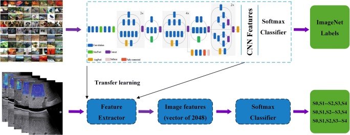
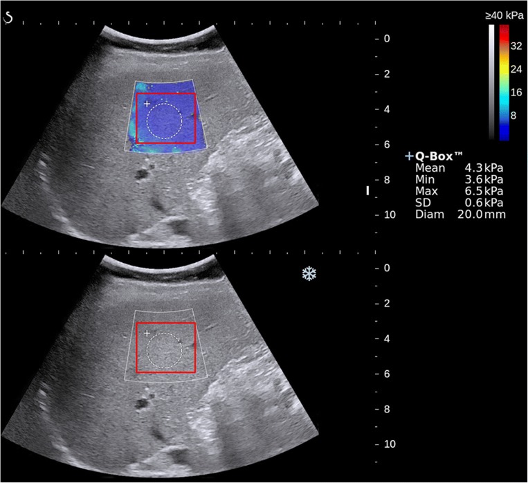
Methods: Totally 466 patients undergoing partial hepatectomy were enrolled, including 401 with chronic hepatitis B and 65 without fibrosis pathologically. All patients received elastography and got liver stiffness measurement (LSM) 2-3 days before surgery. We proposed a deep convolutional neural network by TL to analyze images of gray scale modality (GM) and elastogram modality (EM). The TL process was used for liver fibrosis classification by Inception-V3 network which pretrained on ImageNet. The diagnostic performance of TL and non-TL was compared. The value of single modalities, including GM and EM alone, and multimodalities, including GM + LSM and GM + EM, was evaluated and compared with that of LSM and serological indexes. Receiver operating characteristic curve analysis was performed to calculate the optimal area under the curve (AUC) for classifying fibrosis of S4, ≥ S3, and ≥ S2.
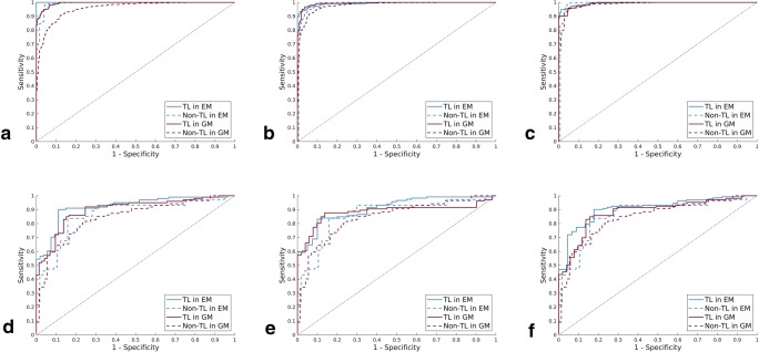
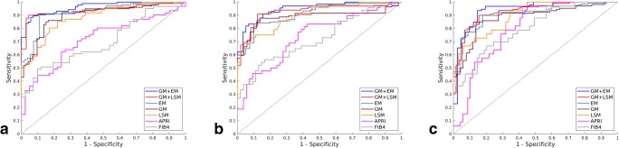
Results: TL in GM and EM demonstrated higher diagnostic accuracy than non-TL, with significantly higher AUCs (all p < .01). Single-modal GM and EM both performed better than LSM and serum indexes (all p < .001). Multimodal GM + EM was the most accurate prediction model (AUCs are 0.950, 0.932, and 0.930 for classifying S4, ≥ S3, and ≥ S2, respectively) compared with GM + LSM, GM and EM alone, LSM, and biomarkers (all p < .05).
Conclusions: Liver fibrosis can be staged by a transfer learning modal based on the combination of gray scale and elastogram ultrasound images, with excellent performance.
Key points: • Transfer learning consists in applying to a specific deep learning algorithm that pretrained on another relevant problem, expected to reduce the risk of overfitting due to insufficient medical images. • Liver fibrosis can be staged by transfer learning radiomics with excellent performance. • The most accurate prediction model of transfer learning by Inception-V3 network is the combination of gray scale and elastogram ultrasound images.
Keywords: Deep learning; Elasticity imaging techniques; Hepatitis B; Liver cirrhosis.
Conflict of interest statement
The authors of this manuscript declare no relationships with any companies whose products or services may be related to the subject matter of the article.
Figures

Similar articles
-
Multiparametric ultrasomics of significant liver fibrosis: A machine learning-based analysis.Eur Radiol. 2019 Mar;29(3):1496-1506. doi: 10.1007/s00330-018-5680-z. Epub 2018 Sep 3. Eur Radiol. 2019. PMID: 30178143 Free PMC article.
-
Comparing radiomics models with different inputs for accurate diagnosis of significant fibrosis in chronic liver disease.Eur Radiol. 2021 Nov;31(11):8743-8754. doi: 10.1007/s00330-021-07934-6. Epub 2021 Apr 21. Eur Radiol. 2021. PMID: 33881568
-
Identifying liver cirrhosis in patients with chronic hepatitis B: an interpretable machine learning algorithm based on LSM.Ann Med. 2025 Dec;57(1):2477294. doi: 10.1080/07853890.2025.2477294. Epub 2025 Mar 19. Ann Med. 2025. PMID: 40104981 Free PMC article.
-
Assessment of biopsy-proven liver fibrosis by two-dimensional shear wave elastography: An individual patient data-based meta-analysis.Hepatology. 2018 Jan;67(1):260-272. doi: 10.1002/hep.29179. Epub 2017 Nov 15. Hepatology. 2018. PMID: 28370257 Free PMC article. Review.
-
Noninvasive diagnosis of liver cirrhosis: qualitative and quantitative imaging biomarkers.Abdom Radiol (NY). 2024 Jun;49(6):2098-2115. doi: 10.1007/s00261-024-04225-8. Epub 2024 Feb 19. Abdom Radiol (NY). 2024. PMID: 38372765 Review.
Cited by
-
Detection of liver cirrhosis in standard T2-weighted MRI using deep transfer learning.Eur Radiol. 2021 Nov;31(11):8807-8815. doi: 10.1007/s00330-021-07858-1. Epub 2021 May 11. Eur Radiol. 2021. PMID: 33974149 Free PMC article.
-
Advances in Deep Learning-Based Medical Image Analysis.Health Data Sci. 2021 May 19;2021:8786793. doi: 10.34133/2021/8786793. eCollection 2021. Health Data Sci. 2021. PMID: 38487506 Free PMC article. Review.
-
Conventional and artificial intelligence-based imaging for biomarker discovery in chronic liver disease.Hepatol Int. 2022 Jun;16(3):509-522. doi: 10.1007/s12072-022-10303-0. Epub 2022 Feb 9. Hepatol Int. 2022. PMID: 35138551 Free PMC article. Review.
-
Multi-site, multi-vendor development and validation of a deep learning model for liver stiffness prediction using abdominal biparametric MRI.Eur Radiol. 2025 Jul;35(7):4362-4373. doi: 10.1007/s00330-024-11312-3. Epub 2025 Jan 9. Eur Radiol. 2025. PMID: 39779515
-
Artificial intelligence in liver imaging: methods and applications.Hepatol Int. 2024 Apr;18(2):422-434. doi: 10.1007/s12072-023-10630-w. Epub 2024 Feb 20. Hepatol Int. 2024. PMID: 38376649 Review.
References
-
- Oshiro H, Itoi T, Iwatsuka K, et al. Liver fibrosis: noninvasive assessment using supersonic shear imaging and FIB4 index in patients with non-alcoholic fatty liver disease. J Med Ultrason (2001) 2017;45:243–249. - PubMed
-
- (1994) Intraobserver and interobserver variations in liver biopsy interpretation in patients with chronic hepatitis C. The French METAVIR Cooperative Study Group. Hepatology 20:15–20 - PubMed
MeSH terms
Substances
Grants and funding
LinkOut - more resources
Full Text Sources
Other Literature Sources
Medical
