

Therapeutic Role of Neuregulin 1 Type III in SOD1-Linked Amyotrophic Lateral Sclerosis
- PMID: 31965551
- PMCID: PMC7609630
- DOI: 10.1007/s13311-019-00811-7
Therapeutic Role of Neuregulin 1 Type III in SOD1-Linked Amyotrophic Lateral Sclerosis
Abstract
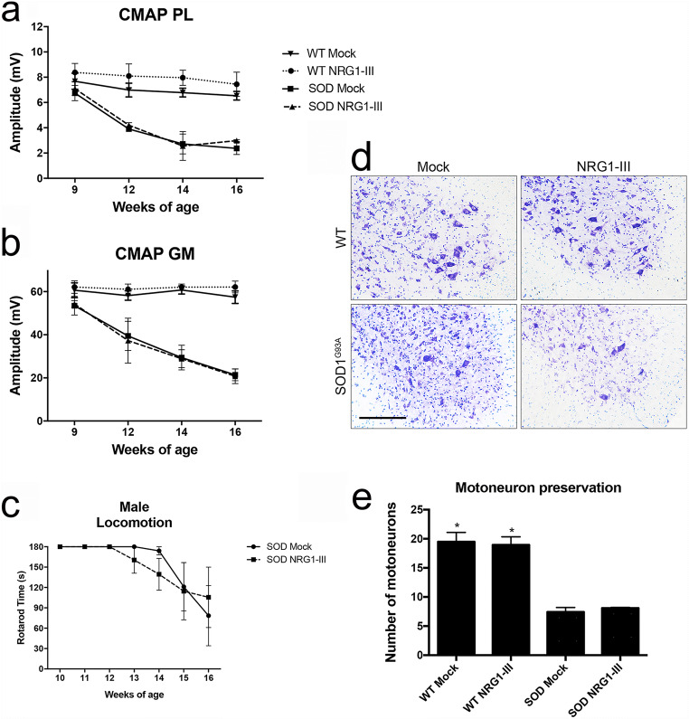
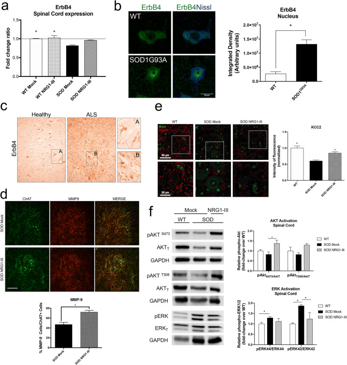
Amyotrophic lateral sclerosis (ALS) is a devastating motoneuron (Mn) disease without effective cure currently available. Death of MNs in ALS is preceded by failure of neuromuscular junctions and axonal retraction. Neuregulin 1 (NRG1) is a neurotrophic factor highly expressed in MNs and neuromuscular junctions that support axonal and neuromuscular development and maintenance. NRG1 and its ErbB receptors are involved in ALS. Reduced NRG1 expression has been found in ALS patients and in the ALS SOD1G93A mouse model; however, the expression of the isoforms of NRG1 and its receptors is still controversial. Due to the reduced levels of NRG1 type III (NRG1-III) in the spinal cord of ALS patients, we used gene therapy based on intrathecal administration of adeno-associated virus to overexpress NRG1-III in SOD1G93A mice. The mice were evaluated from 9 to 16 weeks of age by electrophysiology and rotarod tests. At 16 weeks, samples were harvested for histological and molecular analyses. Our results indicate that overexpression of NRG1-III is able to preserve neuromuscular function of the hindlimbs, improve locomotor performance, increase the number of surviving MNs, and reduce glial reactivity in the treated female SOD1G93A mice. Furthermore, the NRG1-III/ErbB4 axis appears to regulate MN excitability by modulating the chloride transporter KCC2 and reduces the expression of the MN vulnerability marker MMP-9. However, NRG1-III did not have a significant effect on male mice, indicating relevant sex differences. These findings indicate that increasing NRG1-III at the spinal cord is a promising approach for promoting MN protection and functional improvement in ALS.
Keywords: Amyotrophic lateral sclerosis; ErbB receptor; motoneuron disease; motor system; mouse; neuregulin.
Figures


Similar articles
-
Persistent NRG1 Type III Overexpression in Spinal Motor Neurons Has No Therapeutic Effect on ALS-Related Pathology in SOD1G93A Mice.Neurotherapeutics. 2023 Oct;20(6):1820-1834. doi: 10.1007/s13311-023-01424-x. Epub 2023 Sep 21. Neurotherapeutics. 2023. PMID: 37733208 Free PMC article.
-
Gene therapy for overexpressing Neuregulin 1 type I in skeletal muscles promotes functional improvement in the SOD1G93A ALS mice.Neurobiol Dis. 2020 Apr;137:104793. doi: 10.1016/j.nbd.2020.104793. Epub 2020 Feb 4. Neurobiol Dis. 2020. PMID: 32032731
-
Neuregulin 1 confers neuroprotection in SOD1-linked amyotrophic lateral sclerosis mice via restoration of C-boutons of spinal motor neurons.Acta Neuropathol Commun. 2016 Feb 18;4:15. doi: 10.1186/s40478-016-0286-7. Acta Neuropathol Commun. 2016. PMID: 26891847 Free PMC article.
-
Neuregulin-1 and ALS19 (ERBB4): at the crossroads of amyotrophic lateral sclerosis and cancer.BMC Med. 2024 Feb 19;22(1):74. doi: 10.1186/s12916-024-03293-3. BMC Med. 2024. PMID: 38369520 Free PMC article. Review.
-
ALS genetic modifiers that increase survival of SOD1 mice and are suitable for therapeutic development.Prog Neurobiol. 2011 Oct;95(2):133-48. doi: 10.1016/j.pneurobio.2011.07.009. Epub 2011 Jul 26. Prog Neurobiol. 2011. PMID: 21816207 Review.
Cited by
-
Extracellular Vesicles in Serum and Central Nervous System Tissues Contain microRNA Signatures in Sporadic Amyotrophic Lateral Sclerosis.Front Mol Neurosci. 2021 Oct 29;14:739016. doi: 10.3389/fnmol.2021.739016. eCollection 2021. Front Mol Neurosci. 2021. PMID: 34776863 Free PMC article.
-
Persistent NRG1 Type III Overexpression in Spinal Motor Neurons Has No Therapeutic Effect on ALS-Related Pathology in SOD1G93A Mice.Neurotherapeutics. 2023 Oct;20(6):1820-1834. doi: 10.1007/s13311-023-01424-x. Epub 2023 Sep 21. Neurotherapeutics. 2023. PMID: 37733208 Free PMC article.
-
Gene Therapy Overexpressing Neuregulin 1 Type I in Combination With Neuregulin 1 Type III Promotes Functional Improvement in the SOD1G93A ALS Mice.Front Neurol. 2021 Sep 22;12:693309. doi: 10.3389/fneur.2021.693309. eCollection 2021. Front Neurol. 2021. PMID: 34630277 Free PMC article.
-
Disease propagation in amyotrophic lateral sclerosis (ALS): an interplay between genetics and environment.J Neuroinflammation. 2020 Jun 6;17(1):175. doi: 10.1186/s12974-020-01849-7. J Neuroinflammation. 2020. PMID: 32505190 Free PMC article. Review.
-
Identifying dysregulated regions in amyotrophic lateral sclerosis through chromatin accessibility outliers.HGG Adv. 2024 Jul 18;5(3):100318. doi: 10.1016/j.xhgg.2024.100318. Epub 2024 Jun 13. HGG Adv. 2024. PMID: 38872308 Free PMC article.
References
-
- Rosen DR, Siddique T, Patterson D, et al. Mutations in Cu/Zn superoxide dismutase gene are associated with familial amyotrophic lateral sclerosis. Nature. 1993;364:362. - PubMed
-
- Kabashi E, Valdmanis PN, Dion P, et al. TARDBP mutations in individuals with sporadic and familial amyotrophic lateral sclerosis. Nat Genet. 2008;40:572–574. - PubMed
-
- Yokoseki A, Shiga A, Tan CF, et al. TDP-43 mutation in familial amyotrophic lateral sclerosis. Ann Neurol. 2008;63:538–542. - PubMed
Publication types
MeSH terms
Substances
LinkOut - more resources
Full Text Sources
Medical
Research Materials
Miscellaneous
