Phase-separation in chromatin organization
Affiliations
- PMID: 31965983
- PMCID: PMC9107952
Item in Clipboard
Phase-separation in chromatin organization
J Biosci.
2020.
Abstract
The organization of chromatin into different types of compact versus open states provides a means to fine tune gene regulation. Recent studies have suggested a role for phase-separation in chromatin compaction, raising new possibilities for regulating chromatin compartments. This perspective discusses some specific molecular mechanisms that could leverage such phase-separation processes to control the functions and organization of chromatin.
Figures
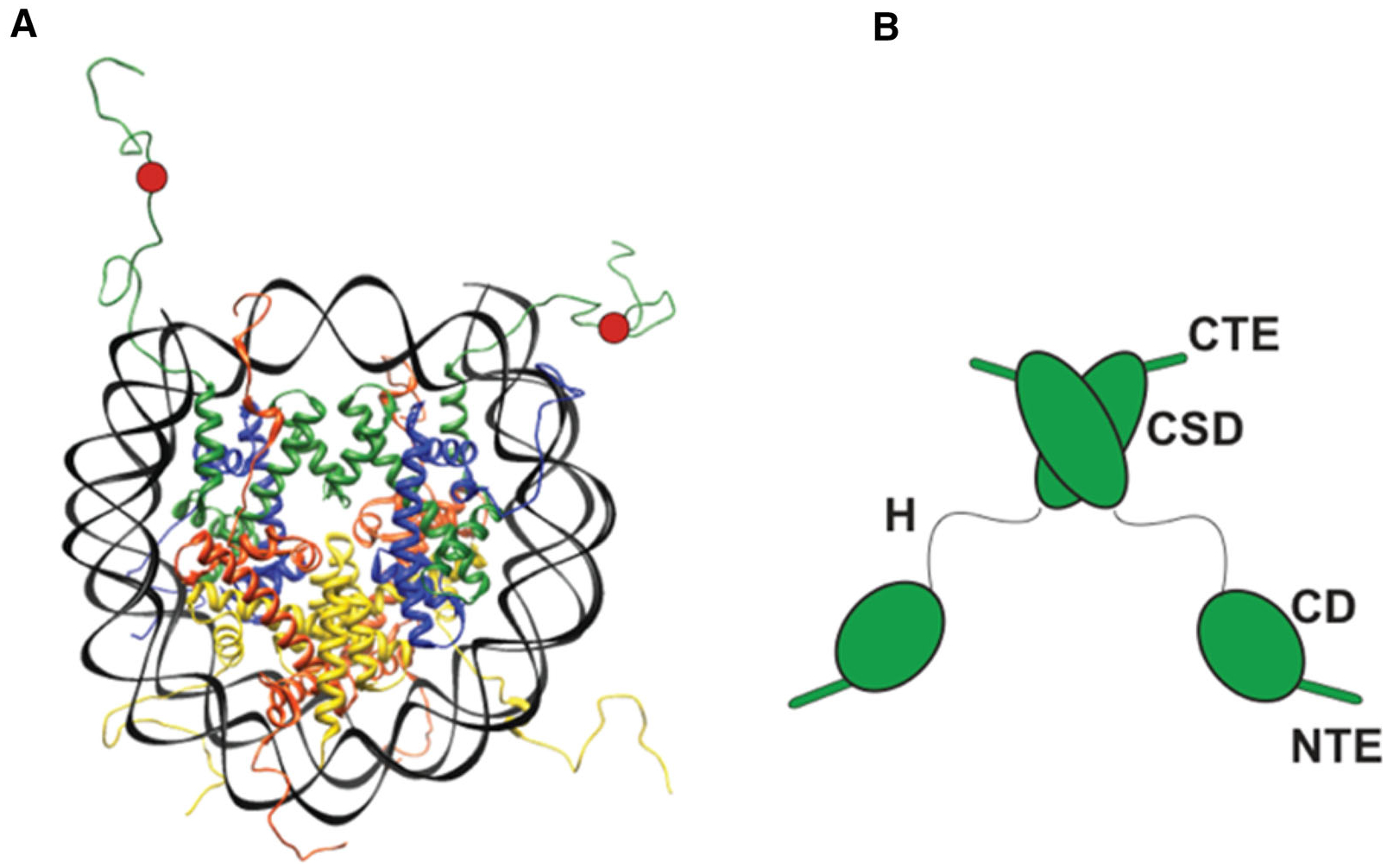
(A) The structure of a nucleosome. DNA is in black. Histone H2A is in red, H2B in yellow, H3 in green and H4 in blue. The H3K9me3 mark is schematically shown as a red circle. (B) Domain diagram of an HP1 dimer. HP1 has two structured domains, the CD, which binds the H3K9me3 mark and the CSD, which forms a dimer that serves as binding interface for various protein ligands. HP1 has three intrinsically disordered regions (IDRs), an N-terminal extension (NTE), a hinge (H) and a C-terminal extension (CTE). Interactions made by the NTE and hinge mediate higher-order HP1 oligomerization.

The schematic shows multiple LLPS droplets across a stretch of the genome and highlights different mechanisms for regulating LLPS in the context of chromatin organization. The droplets are shown in blue and grey. HP1 proteins are shown in green, and other hypothetical chromatin regulators are shown in purple and maroon. Nucleosomes in blue are shown schematically as adopting different shapes to represent different conformations. The H3K9 methyl mark is depicted in red. Thicker arrows represent droplets with higher material strength compared to thinner arrows.
Similar articles
-
The Role of Phase Separation in Heterochromatin Formation, Function, and Regulation.Biochemistry. 2018 May 1;57(17):2540-2548. doi: 10.1021/acs.biochem.8b00401. Epub 2018 Apr 23. Biochemistry. 2018. PMID: 29644850 Free PMC article. Review.
-
Multimodal interactions drive chromatin phase separation and compaction.Proc Natl Acad Sci U S A. 2023 Dec 12;120(50):e2308858120. doi: 10.1073/pnas.2308858120. Epub 2023 Dec 4. Proc Natl Acad Sci U S A. 2023. PMID: 38048471 Free PMC article.
-
Chromatin Compaction Leads to a Preference for Peripheral Heterochromatin.Biophys J. 2020 Mar 24;118(6):1479-1488. doi: 10.1016/j.bpj.2020.01.034. Epub 2020 Feb 4. Biophys J. 2020. PMID: 32097622 Free PMC article.
-
HP1 reshapes nucleosome core to promote phase separation of heterochromatin.Nature. 2019 Nov;575(7782):390-394. doi: 10.1038/s41586-019-1669-2. Epub 2019 Oct 16. Nature. 2019. PMID: 31618757 Free PMC article.
-
Chromatin regulation of plant development.Curr Opin Plant Biol. 2003 Feb;6(1):20-8. doi: 10.1016/s1369526602000079. Curr Opin Plant Biol. 2003. PMID: 12495747 Review.
Cited by
-
'RNA modulation of transport properties and stability in phase-separated condensates.Biophys J. 2021 Dec 7;120(23):5169-5186. doi: 10.1016/j.bpj.2021.11.003. Epub 2021 Nov 9. Biophys J. 2021. PMID: 34762868 Free PMC article.
-
OpenABC Enables Flexible, Simplified, and Efficient GPU Accelerated Simulations of Biomolecular Condensates.bioRxiv [Preprint]. 2023 Apr 21:2023.04.19.537533. doi: 10.1101/2023.04.19.537533. bioRxiv. 2023. Update in: PLoS Comput Biol. 2023 Sep 11;19(9):e1011442. doi: 10.1371/journal.pcbi.1011442. PMID: 37131742 Free PMC article. Updated. Preprint.
-
Polymer model integrates imaging and sequencing to reveal how nanoscale heterochromatin domains influence gene expression.Nat Commun. 2025 Apr 23;16(1):3816. doi: 10.1038/s41467-025-59001-z. Nat Commun. 2025. PMID: 40268925 Free PMC article.
-
Chromatin Liquid-Liquid Phase Separation (LLPS) Is Regulated by Ionic Conditions and Fiber Length.Cells. 2022 Oct 6;11(19):3145. doi: 10.3390/cells11193145. Cells. 2022. PMID: 36231107 Free PMC article.
-
Molecular principles of Piwi-mediated cotranscriptional silencing through the dimeric SFiNX complex.Genes Dev. 2021 Mar 1;35(5-6):392-409. doi: 10.1101/gad.347989.120. Epub 2021 Feb 11. Genes Dev. 2021. PMID: 33574069 Free PMC article.
References
-
- Grewal SI and Elgin SC 2002. Heterochromatin new possibilities for the inheritance of structure. Curr. Opin. Genet. Dev 12 178–187 - PubMed