Review
. 2020:45:21.

TET methylcytosine oxidases: new insights from a decade of research

Affiliations
Review

TET methylcytosine oxidases: new insights from a decade of research

Chan-Wang J Lio et al. J Biosci. 2020.

Abstract

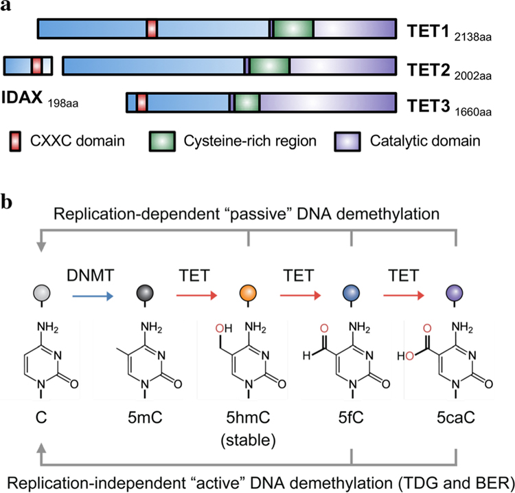
In mammals, DNA methyltransferases transfer a methyl group from S-adenosylmethionine to the 5 position of cytosine in DNA. The product of this reaction, 5-methylcytosine (5mC), has many roles, particularly in suppressing transposable and repeat elements in DNA. Moreover, in many cellular systems, cell lineage specification is accompanied by DNA demethylation at the promoters of genes expressed at high levels in the differentiated cells. However, since direct cleavage of the C-C bond connecting the methyl group to the 5 position of cytosine is thermodynamically disfavoured, the question of whether DNA methylation was reversible remained unclear for many decades. This puzzle was solved by our discovery of the TET (Ten- Eleven Translocation) family of 5-methylcytosine oxidases, which use reduced iron, molecular oxygen and the tricarboxylic acid cycle metabolite 2-oxoglutarate (also known as a-ketoglutarate) to oxidise the methyl group of 5mC to 5-hydroxymethylcytosine (5hmC) and beyond. TET-generated oxidised methylcytosines are intermediates in at least two pathways of DNA demethylation, which differ in their dependence on DNA replication. In the decade since their discovery, TET enzymes have been shown to have important roles in embryonic development, cell lineage specification, neuronal function and cancer. We review these findings and discuss their implications here.


Conflict of interest statement

Conflict of interest AR is on the scientific advisory board of Cambridge Epigenetix (Cambridge, UK). The other authors declare no competing interests.

Figures

Figure 1.
Figure 1.
Ten-Eleven Translocation (TET) proteins and DNA modification. (a) TET family proteins. Mammalian genomes encode three members of the TET/JBP family: TET1, TET2, and TET3. The diagram depicts the domain structures and the length in amino acids (aa) of human TET proteins. The CXXC domains of TET1 and TET3 (red) bind unmethylated CpG sequences in DNA. Note that during evolution, the CXXC domain of primordial TET2 was separated from the TET2 catalytic domain due to chromosomal inversion and evolved as a different gene (IDAX or CXXC4). All three TET proteins contain cysteine-rich domains (green) followed by a C-terminal catalytic domain (purple). (b) TET-mediated DNA modifications and demethylation. DNA methyltransferases (DNMT) methylate unmodified cytosines (C) to yield 5-methylcytosine (5mC). TET proteins can successively oxidize 5mC to 5-hydroxymethylcytosine (5hmC), 5-formylcytosine (5fC), and 5-carboxylcytosine (5caC). Among these three oxidized methylcytosines (oxi-mC), 5hmC is a stable modification and is the most abundant, accounting for ~1–10% of 5mC depending on the cell type, while 5fC and 5caC are ~100-fold and ~1000-fold less abundant than 5hmC. All three oxi-mCs are intermediates for DNA demethylation. During DNA replication, the 5mC at the CpG motif on the template strand pairs with unmodified CpG on the newly synthesized strand, resulting in the hemi-methylated CpG motif. The maintenance methyltransferase complex, DNMT1/UHRF1 binds to the hemi-methylated CpG and rapidly restores methylation on the CpG on the newly synthesized DNA, to restore symmetrical CpG methylation. In contrast, the presence of oxi-mCs in the template strand inhibits the binding of DNMT1/UHRF1 to hemi-modified CpGs, thus preventing methylation of CpGs in the newly synthesized strand. This process is known as ‘passive’ DNA demethylation (top arrows). Additionally, 5fC and 5caC can be recognized and removed by thymine DNA glycosylase (TDG). The abasic site will be repaired by the base-excision repair (BER) system and replaced by an unmodified cytosine, a process termed ‘active’ (replication-independent) DNA demethylation (bottom arrows).
Figure 2.
Figure 2.
Regulation of enhancers by TET proteins. (a) 5hmC levels at enhancers show a strong positive correlation with enhancer activity. The diagram depicts all enhancers in naïve mouse B cells (n=22,539), ranked according to their relative levels of H3-lysine 4-monomethylation (H3K4me1; a mark for most enhancers) and H3-lysine 27-acetylation (H3K27Ac; a mark for enhancer activity). The color indicates the relative enrichment of 5hmC. In general, active enhancers bearing both marks (right) are enriched in 5hmC relative to poised enhancers bearing only the K3K4me1 mark (left). The figure was adapted from Lio et al. (2019) with permission. (b) Working model for TET-mediated enhancer regulation. Pioneer transcription factors (TF1, purple circle) are able to bind to nucleosomes at enhancers and recruit TET proteins. Using 2-oxoglutarate (2OG; also known as alpha-ketoglutarate), reduced iron (Fe(II)) and O2, TET proteins oxidize 5mC into 5hmC at CpG motifs around the enhancer, releasing succinate and CO2. After rounds of DNA replication, the CpG motifs become demethylated and the enhancer becomes more accessible for binding of additional transcription factors (TF2, orange circle).
Figure 3.
Figure 3.
Function of TET proteins in immune system. (a) TET proteins are required for the full potential of enhancers. During T cell development and B cell activation, transcription factors (TFs) recruit TET proteins to the key enhancers that promote the expression of lineage-related genes (Tbx21 and Zbtb7b in T cells; Aicda in B cells) (Tsagaratou et al. 2014; Lio et al. 2019). TET proteins oxidize and demethylate enhancers, augmenting gene expression. In the absence of TET proteins, the inability to demethylate enhancers results in decreased gene expression, potentially by affecting chromatin conformation and the binding of additional transcription factors. (b) TET proteins are required for stable gene expression. A variety of transcription factors recruit TET proteins and assemble at the intronic enhancer (CNS2) of Foxp3, the lineage-defining transcription factor for regulatory T (Treg) cells. This results in the demethylation of ~12 CpGs located in the CNS2 enhancer, a process central to establishing and maintaining the stable expression of Foxp3 (Yue et al. 2016).
Figure 4.
Figure 4.
Proposed model for loss of DNA methylation in heterochromatin of TET-deficient embryonic stem cells. Loss of TET proteins results in relocalization of the de novo methyltransferase DNMT3 proteins, from the heterochromatic compartment to euchromatin regions previously occupied by TET proteins. Potentially, this relocalization contributes both to the heterochromatic DNA hypomethylation and the euchromatin DNA hypermethylation observed in TET-deficient cells. Whether this relocalization also occurs in other systems with TET loss-of-function is still an open question. Adapted from López-Moyado et al. (2019).

References

    1. Altemose N, Miga KH, Maggioni M and Willard HF 2014. Genomic characterization of large heterochromatic gaps in the human genome assembly. PLoS Comput. Biol 10 e1003628. - PMC - PubMed
    1. An J, González-Avalos E, Chawla A, Jeong M, López-Moyado IF, Li W, Goodell MA, Chavez L, Ko M and Rao A 2015. Acute loss of TET function results in aggressive myeloid cancer in mice. Nat. Commun 6 10071. - PMC - PubMed
    1. Aravind L, Abhiman S, and Iyer LM 2011. Natural history of the eukaryotic chromatin protein methylation system. Prog Mol Biol Transl Sci 101 105–176 - PubMed
    1. Aravind L, Balasubramanian S and Rao A 2019. Unusual Activity of a Chlamydomonas TET/JBP Family Enzyme. Biochemistry (in press) - PMC - PubMed
    1. Aravind L and Koonin EV 2001. The DNA-repair protein AlkB, EGL-9, and leprecan define new families of 2-oxoglutarate- and iron-dependent dioxygenases. Genome Biol 2 RESEARCH0007 - PMC - PubMed