

High infiltration of CD68-tumor associated macrophages, predict poor prognosis in Kazakh esophageal cancer patients
- PMID: 31966363
- PMCID: PMC6965792
High infiltration of CD68-tumor associated macrophages, predict poor prognosis in Kazakh esophageal cancer patients
Abstract
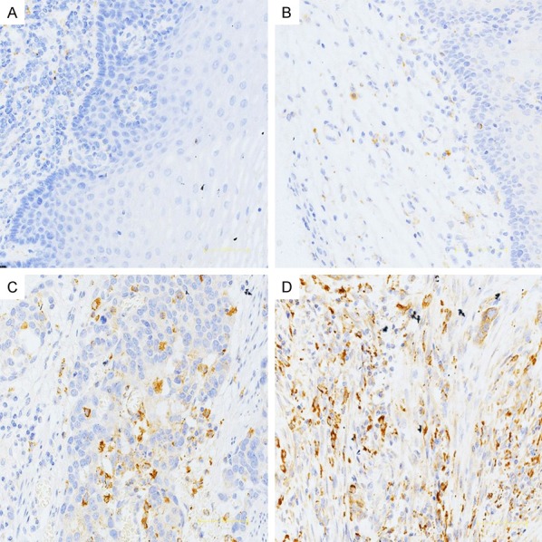
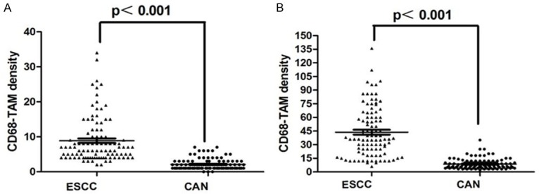
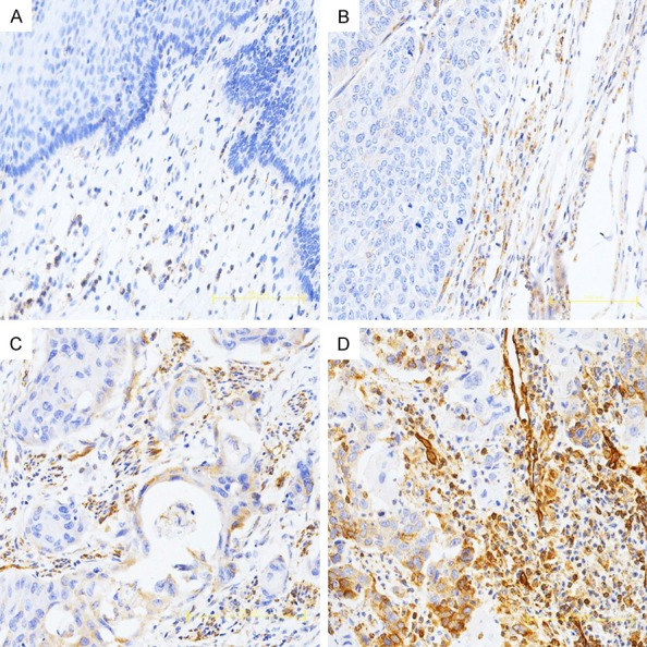
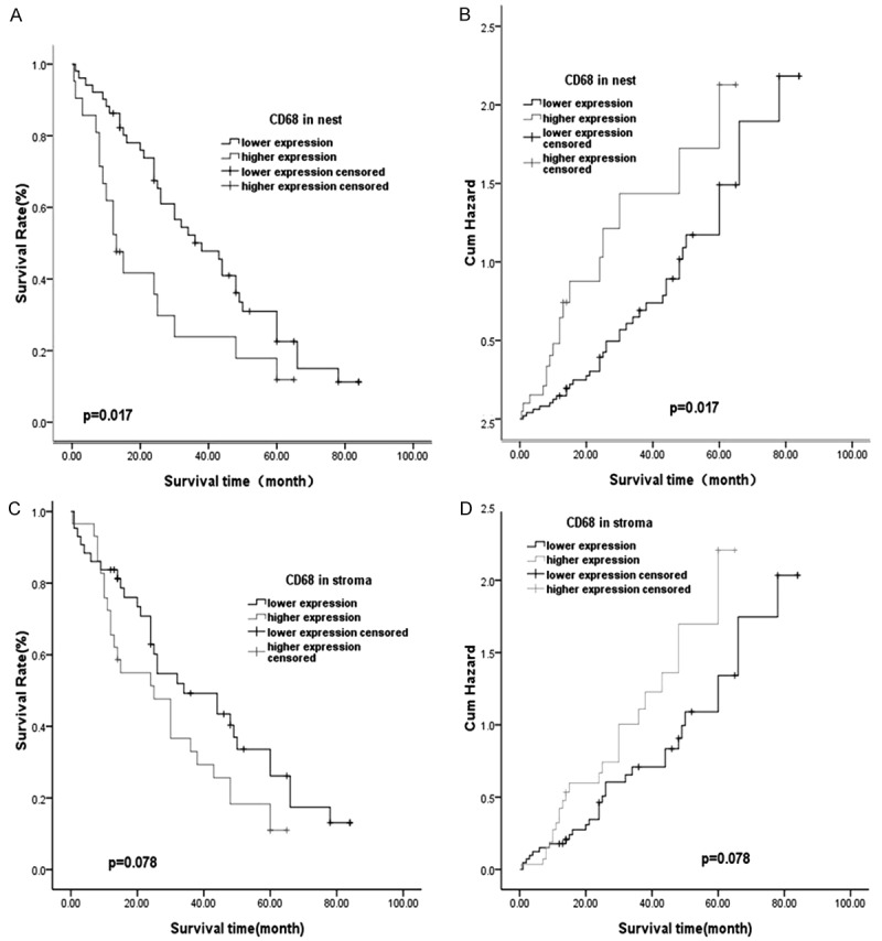
Tumor-associated macrophages (TAMs), the most important immune cells in tumor microenvironment, were reported to play a key role in cancer progression, but the correlation of TAMs and Kazakh esophageal squamous cell carcinoma (ESCC) was still not clear, so we sought to identify the function of TAMs in Kazakh ESCC clinicopathological and prognostic evaluation. CD68 as the TAMs marker, and immunohistochemistry (IHC) was used to quantify the TAMs infiltrated in tumor nest and stroma, the IHC staining was also used to evaluate the expression of MMP-9 in Kazakh ESCCs. The density of CD68-TAMs in ESCCs tumor nest and stromal, were significantly higher than those of CANs (P<0.05). The increasing number of CD68-positive TAMs in tumor nest and stromal were positively associated with tumors lymph node metastasis and clinical stage (P<0.05). The expression of MMP-9 in Kazakh ESCCs was higher than that of CAN tissues (P<0.05). Increased MMP-9 expression in ESCCs was significantly associated with lymph node metastasis and tumor clinical stage (P<0.05). Importantly, the number of CD68-positive TAMs in ESCCs was significantly correlated with the expression of MMP-9 (P<0.05). Furthermore, the survival analyses demonstrated that high-density of CD68-TAMs in tumor nest was positively related to the shorter overall survival time of patients (P<0.05). Increasing numbers of CD68-TAMs promote higher expression of MMP-9 and may play an important role in the occurrence and progression of Kazakh ESCCs, and which could be used as important prognostic markers for Kazakh ESCCs.
Keywords: CD68; Kazakh; MMP-9; Tumor associated macrophages; esophageal squamous cell carcinoma.
IJCEP Copyright © 2017.
Conflict of interest statement
None.
Figures
References
-
- Wang Y, Wu J, Guo W, Sun Q, Chen X, Zhang W, Dong Z, Zhao G. α-Solanine modulates the radiosensitivity of esophageal cancer cells by inducing MicroRNA 138 expression. Cell Physiol Biochem. 2016;39:996–1010. - PubMed
-
- Ayxiam H, Ma H, Ilyar S, Zhang LW, Ablizi A, Batur M, Lu XM. [Metabonomic variation of esophageal cancer within different ethnic groups in Xinjiang, China] . Zhonghua Yu Fang Yi Xue Za Zhi. 2009;43:591–6. - PubMed
-
- Kankuri E, Cholujova D, Comajova M, Vaheri A, Bizik J. Induction of hepatocyte growth factor/scatter factor by fibroblast clustering directly promotes tumor cell invasiveness. Cancer Res. 2005;65:9914–22. - PubMed
-
- Pollard JW. Tumour-educated macrophages promote tumour progression and metastasis. Nat Rev Cancer. 2004;4:71–78. - PubMed
LinkOut - more resources
Full Text Sources
Miscellaneous