. 2017 Jul 1;10(7):7292-7302.
eCollection 2017.

Protective effects of neurotensins on lipopolysaccaride-induced acute lung injury by blocking tachykinin mediated pathway

Affiliations

Protective effects of neurotensins on lipopolysaccaride-induced acute lung injury by blocking tachykinin mediated pathway

Shuiqiao Fu et al. Int J Clin Exp Pathol. .

Abstract

Neurotensin, a bioactive tridecapeptide, has been shown to regulate inflammatory process in lung tissues. However, the effect of neurotensin on LPS-induced lung injury and underlying detailed molecular mechanisms has not been studied. The aim of present study is to investigate the effect of neurotensin on LPS-induced acute lung injury in mice. Mice were treated with LPS intratracheally to induce acute lung injury. 1 hour after ALI induction, and then mice were treated with neurotensins (NTs) (20 mg/kg, 40 mg/kg, and 80 mg/kg) via tail vein injection. Next, the severity of lung injury, MPO activity, neutrophils infiltration, lung edema, protein and pro-inflammatory cytokines concentration in BALF were determined to evaluate the effect of Nts on ALI. Additionally, the expression of tachykinins receptors, including NK1, NK2, and NK3 and the production of IL-8, COX-2, and PGE2 mediated by tachykinins-tachykinins receptors pathway were determined to investigate the blocking effect of Nts on tachykinins and its receptors pathway. Neurotensins treatment significantly decreased the lung edema and the infiltration of inflammatory cells into lung tissue caused by LPS induction. Meanwhile, the elevation of pro-inflammatory cytokines and chemokine in BALF was dramatically reduced by neurotensins treatment. Furthermore, neurotensins could interact with tachykinins receptors and block the inflammatory responses activated by tachykinins pathways. In summary, neurotensins has a potentially protective effect on LPS-induced acute lung injury through the interaction with tachykinins receptors and subsequently blocking the inflammatory responses induced by activation of tachykinins pathway.

Keywords: COX-2; Neurotensin; PGE2; acute lung injury; inflammation; tachykinins.


Conflict of interest statement

None.

Figures

Figure 1
Figure 1
LPS induction increased the expression of tachykinins receptors in vivo. A: The mRNA level of tachykinins receptors were determined by Real-time PCR assay; B: The protein levels of tachykinins receptors were measured by western blot analysis methods. Results are expressed as mean ± SD. *P<0.05 compared with the control group.
Figure 2
Figure 2
The effects of Nts on the lung histopathological changes. The lung tissues isolated from differentially treated ALI mice were stained with hematoxylin and eosin (×200). A. The lung section from of a control mice; B. The lung section from of LPS-induced ALI mice; C. The lung section from of 20 mg/kg Nts treated ALI mice; D. The lung section from 40 mg/kg Nts treated ALI mice; E. The lung section from 80 mg/kg Nts treated ALI mice. F. The lung injury index of differentially treated ALI mice. Results are expressed as mean ± SD. *P<0.05 compared with the control group; #P<0.05 compared with the LPS-induced ALI group.
Figure 3
Figure 3
Effects of Nts on MPO activity (A), neutrophils infiltration (B), pulmonary edema (C) and protein leakage in BALF (D). Results are expressed as mean ± SD. *P<0.05 compared with the control group; #P<0.05 compared with the LPS-induced ALI group.
Figure 4
Figure 4
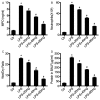
Effect of Nts on the production of inflammatory cytokines and chemokine in BALF from differentially treated ALI mice. A: The level of TNF-α in BALF from differentially treated ALI mice; B: The level of IL-6 in BALF from differentially treated ALI mice; C: The level of IL-1β in BALF from differentially treated ALI mice; D: The level of MCP-1 in BALF from differentially treated ALI mice. Results are expressed as mean ± SD. *P<0.05 compared with the control group; #P<0.05 compared with the LPS-induced ALI group.
Figure 5
Figure 5
Nts treatment blocks the expression of IL-8 mediated by NR1 in vitro. A: The mRNA level of IL-8 in differentially treated RAW264.7 cells; B: The protein level of IL-8 in differentially treated RAW264.7 cells. Results are expressed as mean ± SD. *P<0.05.
Figure 6
Figure 6
Effect of Nts on the expression of COX-2 and PGE2 in vivo and in vitro. A: The protein level of COX-2 in BALF from differentially treated ALI mice; B: The protein level of PGE2 in BALF form differentially treated ALI mice; C: The protein level of COX-2 in differentially treated RAW264.7 cells in vitro; D: The protein level of PGE2 in differentially treated RAW264.7 cells in vitro. Results are expressed as mean ± SD. *P<0.05 compared with the control group; #P<0.05 compared with the LPS-induced ALI group; +, Represent P<0.05.
Figure 7
Figure 7
The effect of Nts on LPS-induced mortality in mice. The mice were treated by LPS with or without Nts (or CP96345) treatment. The survival rates were observed for 1, 24, 48, 72, 96, and 120 h. *P<0.05 compared with the control group; #P<0.05 compared with the LPS-induced ALI group.

References

    1. Matthay MA, Ware LB, Zimmerman GA. The acute respiratory distress syndrome. J Clin Invest. 2012;122:2731–2740. - PMC - PubMed
    1. Ware LB, Matthay MA. The acute respiratory distress syndrome. N Engl J Med. 2000;342:1334–1349. - PubMed
    1. Mei SH, McCarter SD, Deng Y, Parker CH, Liles WC, Stewart DJ. Prevention of LPS-induced acute lung injury in mice by mesenchymal stem cells overexpressing angiopoietin 1. PLoS Med. 2007;4:e269. - PMC - PubMed
    1. Matute-Bello G, Frevert CW, Martin TR. Animal models of acute lung injury. Am J Physiol Lung Cell Mol Physiol. 2008;295:L379–399. - PMC - PubMed
    1. Cannon JG, Tompkins RG, Gelfand JA, Michie HR, Stanford GG, van der Meer JW, Endres S, Lonnemann G, Corsetti J, Chernow B, et al. Circulating interleukin-1 and tumor necrosis factor in septic shock and experimental endotoxin fever. J Infect Dis. 1990;161:79–84. - PubMed

LinkOut - more resources