Florid cystic endosalpingiosis associated with a retroperitoneal leiomyoma mimicking malignancy: a case report
- PMID: 31966902
- PMCID: PMC6965923
Florid cystic endosalpingiosis associated with a retroperitoneal leiomyoma mimicking malignancy: a case report
Abstract
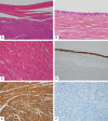
Florid cystic endosalpingiosis (FCE) is a rare type of endosalpingiosis that presents as a mass-like lesion. Here we report an unusual case of FCE associated with a retroperitoneal leiomyoma. A 46-year old female presented with a palpable abdominal mass. A pelvic CT revealed a 23.5×16.3×9.4 cm sized multilocular cystic and solid mass in the retroperitoneum. Surgical excision of the mass was performed. Microscopically, the cystic spaces were lined by a single layer of ciliated tubal epithelium. The solid areas consisted of thick bundles of spindle cells. There were no cytologic atypia, mitosis or necrosis. The spindle cells were positive for actin and desmin, and were negative for c-kit, CD34, S100 and HMB-45, confirming the diagnosis of FCE associated with retroperitoneal leiomyoma.
Keywords: Florid cystic endosalpingiosis; endosalpingiosis; leiomyoma; mimicker; retroperitoneal mass.
IJCEP Copyright © 2017.
Conflict of interest statement
None.
Figures


Similar articles
-
Intramural florid cystic endosalpingiosis of the uterus: a case report and review of the literature.Taiwan J Obstet Gynecol. 2015 Feb;54(1):75-7. doi: 10.1016/j.tjog.2014.11.011. Taiwan J Obstet Gynecol. 2015. PMID: 25675925 Review.
-
Intramural florid cystic endosalpingiosis of the uterus after menopause.Pol J Pathol. 2018;69(3):321-324. doi: 10.5114/pjp.2018.79554. Pol J Pathol. 2018. PMID: 30509061
-
Tumor-like cystic endosalpingiosis in the myometrium: a case report and a review of the literature.Turk Patoloji Derg. 2014;30(2):145-8. doi: 10.5146/tjpath.2013.01176. Turk Patoloji Derg. 2014. PMID: 24101353 Review.
-
Hysteroscopic Findings Related with the Assessment and Treatment of Uterine Florid Cystic Endosalpingiosis: A Case Report and Review of All the Published Cases.Acta Med Port. 2021 Dec 2;34(12):868-873. doi: 10.20344/amp.14292. Epub 2020 Sep 29. Acta Med Port. 2021. PMID: 32991276
-
Retroperitoneal lymphangioleiomyomatosis associated with endosalpingiosis.APMIS. 2007 Dec;115(12):1460-5. doi: 10.1111/j.1600-0463.2007.00668.x. APMIS. 2007. PMID: 18184421
Cited by
-
Intraoperative Appearance of Endosalpingiosis: A Single-Center Experience of Laparoscopic Findings and Systematic Review of Literature.J Clin Med. 2022 Nov 27;11(23):7006. doi: 10.3390/jcm11237006. J Clin Med. 2022. PMID: 36498581 Free PMC article.
-
[Multicentric Florid Cystic Endosalpingiosis in Different Anatomical Spaces: A Case Report].Taehan Yongsang Uihakhoe Chi. 2021 Mar;82(2):481-486. doi: 10.3348/jksr.2020.0098. Epub 2020 Dec 30. Taehan Yongsang Uihakhoe Chi. 2021. PMID: 36238742 Free PMC article. Korean.
-
Giant retroperitoneal myolipoma mimicking liposarcoma: report of a resected case and review of the literature.Int Cancer Conf J. 2024 Feb 5;13(2):144-152. doi: 10.1007/s13691-024-00655-9. eCollection 2024 Apr. Int Cancer Conf J. 2024. PMID: 38524654 Free PMC article.
References
-
- Clement PB, Young RH. Florid cystic endosalpingiosis with tumor-like manifestations: a report of four cases including the first reported cases of transmural endosalpingiosis of the uterus. Am J Surg Pathol. 1999;23:166–175. - PubMed
-
- Prentice L, Stewart A, Mohiuddin S, Johnson NP. What is endosalpingiosis? Fertil Steril. 2012;98:942–947. - PubMed
-
- Patonay B, Semer D, Hong H. Florid cystic endosalpingiosis with extensive peritoneal involvement and concurrent bilateral ovarian serous cystadenoma. J Obstet Gynaecol. 2011;31:773–774. - PubMed
-
- Im S, Jung JH, Choi HJ, Kang CS. Intramural florid cystic endosalpingiosis of the uterus: a case report and review of the literature. Taiwan J Obstet Gynecol. 2015;54:75–77. - PubMed
-
- Mulayim B, Serin N, Karatas S, Celik B. Cystic endosalpingiosis of uterus and ovary found on laparoscopy: disease of Haze. J Minim Invasive Gynecol. 2017;24:4–5. - PubMed
Publication types
LinkOut - more resources
Full Text Sources
