Review
. 2020 Apr 12;32(4):223-231.
doi: 10.1093/intimm/dxaa006.

Physiology and pathology of T-cell aging

Affiliations
Review

Physiology and pathology of T-cell aging

Nagahiro Minato et al. Int Immunol. .

Abstract

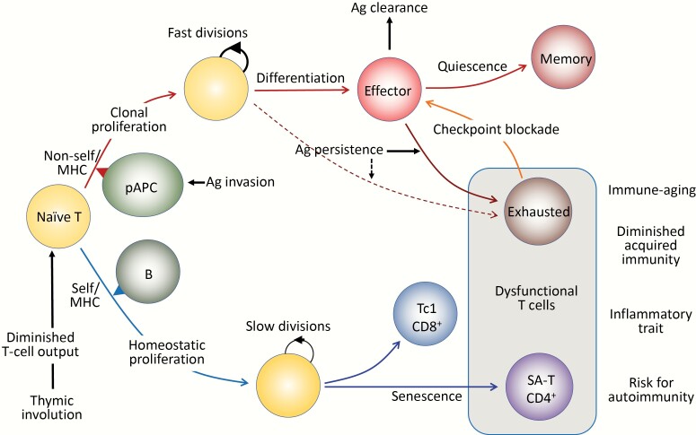
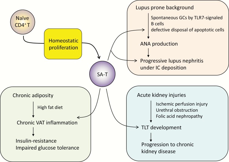
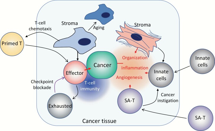
Acquired immune function shows recognizable changes over time with organismal aging. These changes include T-cell dysfunction, which may underlie diminished resistance to infection and possibly various chronic age-associated diseases in the elderly. T-cell dysfunction may occur at distinct stages, from naive cells to the end stages of differentiation during immune responses. The thymus, which generates naive T cells, shows unusually early involution resulting in progressive reduction of T-cell output after adolescence, but peripheral T-cell numbers are maintained through antigen-independent homeostatic proliferation of naive T cells driven by the major histocompatibility complex associated with self-peptides and homeostatic cytokines, retaining the diverse repertoire. However, extensive homeostatic proliferation may lead to the emergence of dysfunctional CD4+ T cells with features resembling senescent cells, termed senescence-associated T (SA-T) cells, which increase and accumulate with age. In situations such as chronic viral infection, T-cell dysfunction may also develop via persistent antigen stimulation, termed exhaustion, preventing possible immunopathology due to excessive immune responses. Exhausted T cells are developed through the effects of checkpoint receptors such as PD-1 and may be reversed with the receptor blockade. Of note, although defective in their regular T-cell antigen-receptor-mediated proliferation, SA-T cells secrete abundant pro-inflammatory factors such as osteopontin, reminiscent of an SA-secretory phenotype. A series of experiments in mouse models indicated that SA-T cells are involved in systemic autoimmunity as well as chronic tissue inflammation following tissue stresses. In this review, we discuss the physiological aspects of T-cell dysfunction associated with aging and its potential pathological involvement in age-associated diseases and possibly cancer.

Keywords: T-cell dysfunction; T-cell exhaustion; age-associated diseases; homeostatic proliferation; senescence-associated T cells.


Figures

Fig. 1.
Fig. 1.
Antigen (Ag)-driven and antigen-independent generation of dysfunctional T cells. (Upper) In response to the optimal TCR stimulation via foreign antigens presented by professional antigen-presenting cells (pAPCs) expressing proper costimulatory molecules, specific naive T cells initiate robust clonal proliferation with fast cell divisions, followed by functional differentiation to various effector cells. As the antigens are cleared, the effector cells may die off, but a portion of them become quiescent and are maintained as central memory T cells. However, when antigen stimulation persists, the effector cells may go into a dysfunctional state via constitutive expression of checkpoint receptors such as PD-1 and LAG3 to prevent immunopathology due to excessive immune responses, called exhausted T cells. The exhausted T cells may also be derived from unique progenitor cells (pre-exhausted T cells). The function of exhausted T cells may be reverted with checkpoint blockade, although these T cells may eventually become refractory. (Lower) Naive T cells that developed through positive selection in the thymus have intrinsic affinity to self-MHCs and are under tonic TCR stimulation, mainly through B cells for CD4+ T cells. Although the tonic TCR signal per se is insufficient for causing proliferation, naive T cells initiate HP with slow cell divisions in the presence of sufficient amounts of homeostatic cytokines (IL-7, IL-15), which are increased in the lymphoid milieu in T-lymphopenic conditions. Thymic involution begins early in life with a progressive reduction of naive T-cell output over time, which increasingly drives HP of naive T cells with age to maintain the size of the T-cell pool in the periphery. Sustained HP of CD8+ T cells leads to the generation of CXCR3+ cells with increased capacity of IFNγ/TNFα production, predisposed to Tc1-type pro-inflammatory T cells. In CD4+ T cells, continuous HP results in the generation of dysfunctional T cells bearing the features resembling cell senescence, termed senescence-associated T (SA-T) cells. The SA-T cells are also characterized by the constitutive expression of PD-1 and LAG3, and partly CD153, although there is so far no evidence that the function is restored by PD-1 checkpoint blockade. SA-T cells are defective in proliferation and regular differentiation in response to optimal TCR stimulation, but these T cells secrete abundant pro-inflammatory cytokines, such as osteopontin and chemokines directed to innate inflammatory cells (Ccl3, 4), reminiscent of SASP. SA-T cells are progressively increased in proportion with age, but in addition, these T cells may remarkably accumulate in the GCs of lymphoid tissues or various other tissues under stresses or insults, predisposing to systemic autoimmunity or age-associated chronic inflammatory diseases.
Fig. 2.
Fig. 2.
Involvement of SA-T cells in systemic autoimmune disease and chronic inflammatory diseases. Detailed explanation may be found in the text. SA-T, senescence-associated CD4+ T cells; GC, germinal center; TLR7, Toll-like receptor 7; ANA, anti-nuclear antibody; IC, immune complex; TLT, tertiary lymphoid tissue; VAT, visceral adipose tissue.
Fig. 3.
Fig. 3.
Schematic representation of T-cell dysfunction and tissue aging in cancer. In cancer tissues, various tissue reactions with often conflicting effects on cancer progression may occur, where tissue stroma cells play important roles. Certain stroma cell types may promote effective recruitment of primed T cells into the tissue via secretion of T-cell chemokines and forming a proper scaffold for their migration. Such T cells at a close enough vicinity to cancer cells proliferate and are activated to become effector cells destroying them. Sustained activation of the T cells, however, may lead to a dysfunctional state, or exhaustion, through checkpoint receptors, such as PD-1, the effects being pronounced when cancer cells express the ligands for checkpoint receptors. The process can be reverted with checkpoint-receptor blockade to resume effective T-cell immunity. The T-cell recruiting activity of stroma cells is radio-sensitive and is also diminished with age possibly through cellular aging. On the other hand, it is also known that stroma cells with potent pro-inflammatory activity are also increased in the tissues of certain types of cancers, often called myofibroblasts with contractile features. Such stroma cells recruit various innate immune cells to cause inflammatory reactions as well as neoangiogenesis and tissue organization. It is also likely that SA-T cells, which secrete abundant chemoattractants for inflammatory cells, such as osteopontin and Ccl3/4 as part of SASP, accumulate, exaggerating the formation of the inflammatory microenvironment around cancer cells. It is reported that osteopontin acts as a potent tumor-instigating factor recruiting myeloid cells directly from bone marrow. Overall, such an inflammatory microenvironment favors the progression of cancer, via damage to normal tissue integrity as well as suppression of T-cell immunity.

References

    1. Franceschi, C., Garagnani, P., Morsiani, C.et al. . 2018. The continuum of aging and age-related diseases: common mechanisms but different rates. Front. Med. (Lausanne) 5:61. - PMC - PubMed
    1. Chung, H. Y., Cesari, M., Anton, S.et al. . 2009. Molecular inflammation: underpinnings of aging and age-related diseases. Ageing Res. Rev. 8:18. - PMC - PubMed
    1. Franceschi, C. and Campisi, J. 2014. Chronic inflammation (inflammaging) and its potential contribution to age-associated diseases. J. Gerontol. A Biol. Sci. Med. Sci. 69(Suppl. 1):S4. - PubMed
    1. Ferrucci, L. and Fabbri, E. 2018. Inflammageing: chronic inflammation in ageing, cardiovascular disease, and frailty. Nat. Rev. Cardiol. 15:505. - PMC - PubMed
    1. van Deursen, J. M. 2014. The role of senescent cells in ageing. Nature 509:439. - PMC - PubMed

Publication types

Substances