Multicenter Study
. 2020 Mar 1;5(3):282-290.
doi: 10.1001/jamacardio.2019.5315.

Association of High-Density Calcified 1K Plaque With Risk of Acute Coronary Syndrome

Affiliations
Multicenter Study

Association of High-Density Calcified 1K Plaque With Risk of Acute Coronary Syndrome

Alexander R van Rosendael et al. JAMA Cardiol. .

Erratum in

Abstract

Importance: Plaque morphologic measures on coronary computed tomography angiography (CCTA) have been associated with future acute coronary syndrome (ACS). However, the evolution of calcified coronary plaques by noninvasive imaging is not known.

Objective: To ascertain whether the increasing density in calcified coronary plaque is associated with risk for ACS.

Design, setting, and participants: This multicenter case-control cohort study included individuals enrolled in ICONIC (Incident Coronary Syndromes Identified by Computed Tomography), a nested case-control study of patients drawn from the CONFIRM (Coronary CT Angiography Evaluation for Clinical Outcomes: An International Multicenter) registry, which included 13 study sites in 8 countries. Patients who experienced core laboratory-verified ACS after baseline CCTA (n = 189) and control individuals who did not experience ACS after baseline CCTA (n = 189) were included. Patients and controls were matched 1:1 by propensity scores for age; male sex; presence of hypertension, hyperlipidemia, and diabetes; family history of premature coronary artery disease (CAD); current smoking status; and CAD severity. Data were analyzed from November 2018 to March 2019.

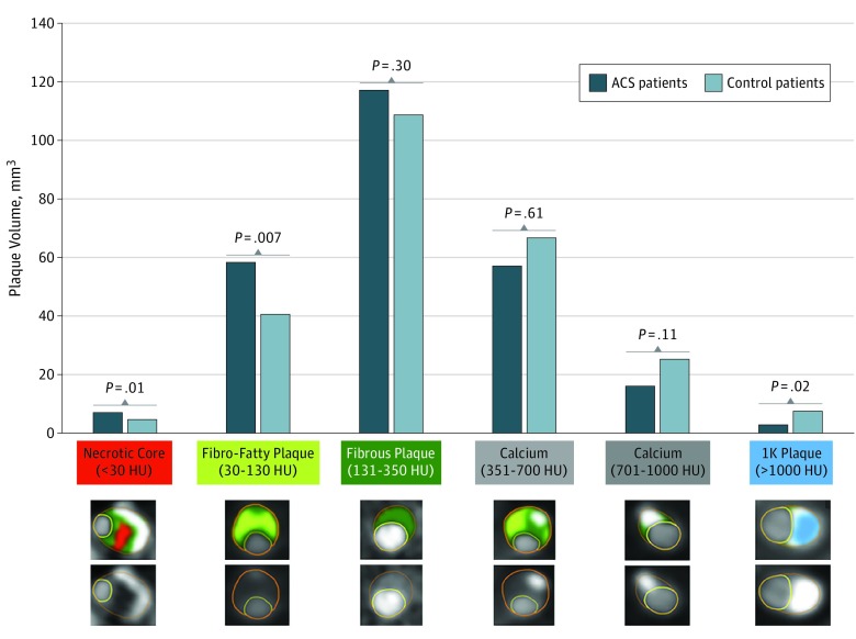
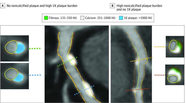
Exposures: Whole-heart atherosclerotic plaque volume was quantitated from all coronary vessels and their branches. For patients who underwent invasive angiography at the time of ACS, culprit lesions were coregistered to baseline CCTA lesions by a blinded independent reader. Low-density plaque was defined as having less than 130 Hounsfield units (HU); calcified plaque, as having more than 350 HU and subcategorized on a voxel-level basis into 3 strata: 351 to 700 HU, 701 to 1000 HU, and more than 1000 HU (termed 1K plaque).

Main outcomes and measures: Association between calcium density and future ACS risk.

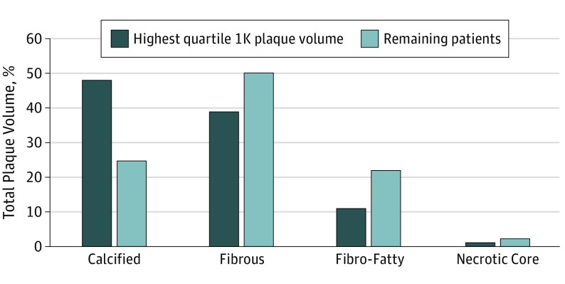
Results: A total of 189 patients and 189 matched controls (mean [SD] age of 59.9 [9.8] years; 247 [65.3%] were male) were included in the analysis and were monitored during a mean (SD) follow-up period of 3.9 (2.5) years. The overall mean (SD) calcified plaque volume (>350 HU) was similar between patients and controls (76.4 [101.6] mm3 vs 99.0 [156.1] mm3; P = .32), but patients who experienced ACS exhibited less 1K plaque (>1000 HU) compared with controls (3.9 [8.3] mm3 vs 9.4 [23.2] mm3; P = .02). Individuals within the highest quartile of 1K plaque exhibited less low-density plaque, as a percentage of total plaque, when compared with patients within the lower 3 quartiles (12.6% [10.4%] vs 24.9% [20.6%]; P < .001). For 93 culprit precursor lesions detected by CCTA, the volume of 1K plaque was lower compared with the maximally stenotic lesion in controls (2.6 [7.2] mm3 vs 7.6 [20.3] mm3; P = .01). The per-patient and per-lesion results were similar between the 2 groups when restricted to myocardial infarction cases.

Conclusions and relevance: Results of this study suggest that, on a per-patient and per-lesion basis, 1K plaque was associated with a lower risk for future ACS and that measurement of 1K plaque may improve risk stratification beyond plaque burden.


Conflict of interest statement

Conflict of Interest Disclosures: Dr Al Hussein Alawamlh reported receiving grants from the National Institutes of Health (NIH) during the conduct of the study. Dr Chinnaiyan reported receiving grants from Heartflow Inc outside the submitted work and being an unpaid medical advisory board member of HeartFlow. Dr Chow reported holding the Saul and Edna Goldfarb Chair in cardiac imaging research, receiving research support from CV Diagnostix and Ausculsciences as well as educational support from TeraRecon Inc, and holding equity interest in GE Healthcare. Dr Cury reported receiving personal fees from Cleerly Inc and GE Healthcare outside the submitted work. Dr Hadamitzky reported receiving nonfinancial support from Siemens Healthineers outside the submitted work. Dr Leipsic reported receiving personal fees from HeartFlow and grants from GE Healthcare outside the submitted work. Dr Pontone reported receiving grants and personal fees from GE Healthcare, grants and personal fees from Bracco, and grants from HeartFlow outside the submitted work. Dr Raff reported receiving grants from Blue Cross Blue Shield of Michigan during the conduct of the study and grants from HeartFlow Inc outside the submitted work. Dr Stuijfzand reported receiving grants from the NIH; Leading Foreign Research Institute Recruitment Program of the National Research Foundation of Korea, Ministry of Science, ICT and Future Planning; and Dalio Institute of Cardiovascular Imaging during the conduct of the study. Dr Samady reported serving on the scientific advisory board of Philips, holding equity interest in Covanos Inc, and receiving research grants from Medtronic, Abbott Vascular, and Philips. Dr Budoff reported receiving grants from GE Healthcare during the conduct of the study. Dr Berman reported receiving software royalties from Cedars-Sinai Medical Center. Dr Bax reported receiving departmental grants and other from Medtronic as well as grants from Biotronik, Boston Scientific, Abbott, Edwards Lifescience, and GE Healthcare during the conduct of the study. Dr Min reported receiving funding from the Dalio Foundation, NIH, and GE Healthcare; serving on the scientific advisory board of Arineta and GE Healthcare; and holding equity interest in Cleerly Inc. No other disclosures were reported.

Figures

Figure 1.
Figure 1.. Whole-Heart Plaque Volume by Composition for Patients With Acute Coronary Syndrome (ACS) and Control Individuals
Per-patient volumes of necrotic core (<30 Hounsfield units [HU]), fibro-fatty (30-130 HU), fibrous (131-350 HU), calcified (351-700 HU), calcified (701-1000 HU), and 1K (>1000 HU) plaques are reported. Patients with ACS reported significantly more necrotic core and fibro-fatty plaques and less 1K plaque compared with control individuals. Cross-sectional examples of the different plaque composition types are displayed by different colors. 1K plaque is shown in blue.
Figure 2.
Figure 2.. Example of a Vessel With 1K Plaque
The artery segment in the left panel (A) shows 2 lesions composed of 1K plaque (ie, plaque with a volume >1000 Hounsfield units [HU]), without noncalcified plaque. Cross-sectional examples are shown with 1K plaque (blue). The artery segment in the right panel (B) shows calcifications between 351 and 1000 HU (gray) intermingled in noncalcified plaque. Two cross-sections show 351- to 1000-HU calcium plaque together with 130- to 350-HU fibrous plaque tissue (green).
Figure 3.
Figure 3.. Plaque Composition in Individuals With a Large 1K Plaque Volume
Individuals with the highest quartile of 1K plaque (ie, plaque with a volume >1000 Hounsfield units [HU]) had relatively more calcified plaque but less fibrous, fibro-fatty, and necrotic core plaques compared with the other 3 quartiles of patients. Absolute volumes and comparisons are provided in eTable 5 in the Supplement.

Comment in

References

    1. Motoyama S, Ito H, Sarai M, et al. . Plaque characterization by coronary computed tomography angiography and the likelihood of acute coronary events in mid-term follow-up. J Am Coll Cardiol. 2015;66(4):337-346. doi:10.1016/j.jacc.2015.05.069 - DOI - PubMed
    1. Chang HJ, Lin FY, Lee SE, et al. . Coronary atherosclerotic precursors of acute coronary syndromes. J Am Coll Cardiol. 2018;71(22):2511-2522. doi:10.1016/j.jacc.2018.02.079 - DOI - PMC - PubMed
    1. Mori H, Torii S, Kutyna M, Sakamoto A, Finn AV, Virmani R. Coronary artery calcification and its progression: what does it really mean? JACC Cardiovasc Imaging. 2018;11(1):127-142. doi:10.1016/j.jcmg.2017.10.012 - DOI - PubMed
    1. Criqui MH, Denenberg JO, Ix JH, et al. . Calcium density of coronary artery plaque and risk of incident cardiovascular events. JAMA. 2014;311(3):271-278. doi:10.1001/jama.2013.282535 - DOI - PMC - PubMed
    1. Min JK, Dunning A, Lin FY, et al. . Rationale and design of the CONFIRM (Coronary CT Angiography Evaluation for Clinical Outcomes: An International Multicenter) Registry. J Cardiovasc Comput Tomogr. 2011;5(2):84-92. doi:10.1016/j.jcct.2011.01.007 - DOI - PubMed

Publication types