

Apoptotic Bodies: Particular Extracellular Vesicles Involved in Intercellular Communication
- PMID: 31968627
- PMCID: PMC7168913
- DOI: 10.3390/biology9010021
Apoptotic Bodies: Particular Extracellular Vesicles Involved in Intercellular Communication
Abstract
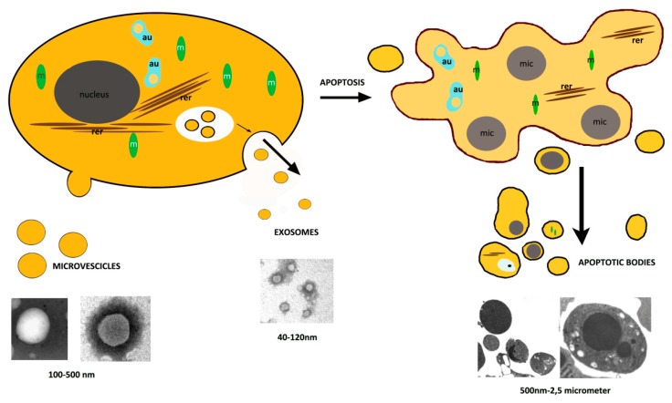
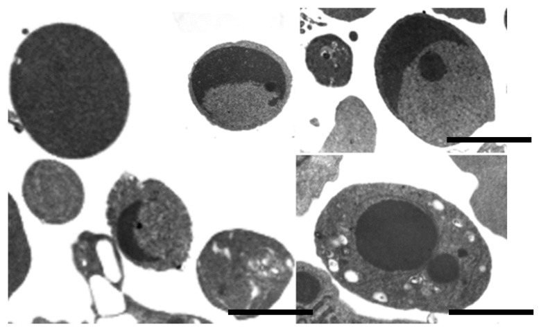
In the last decade, a new method of cell-cell communication mediated by membranous extracellular vesicles (EVs) has emerged. EVs, including exosomes, microvesicles, and apoptotic bodies (ApoBDs), represent a new and important topic, because they are a means of communication between cells and they can also be involved in removing cellular contents. EVs are characterized by differences in size, origin, and content and different types have different functions. They appear as membranous sacs released by a variety of cells, in different physiological and patho-physiological conditions. Intringuingly, exosomes and microvesicles are a potent source of genetic information carriers between different cell types both within a species and even across a species barrier. New, and therefore still relatively poorly known vesicles are apoptotic bodies, on which numerous in-depth studies are needed in order to understand their role and possible function. In this review we would like to analyze their morpho-functional characteristics.
Keywords: apoptotic bodies; extracellular vesicles; intercellular communication; ultrastructure.
Conflict of interest statement
The authors declare no conflict of interest.
Figures

References
-
- Hauser P., Wang S., Didenko V.V. Apoptotic Bodies: Selective Detection in Extracellular Vesicles. Methods Mol. Biol. 2017;1554:193–200. - PubMed
Publication types
LinkOut - more resources
Full Text Sources
Other Literature Sources
