. 2020 Mar 31;94(8):e02151-19.
doi: 10.1128/JVI.02151-19. Print 2020 Mar 31.

Phosphosite Analysis of the Cytomegaloviral mRNA Export Factor pUL69 Reveals Serines with Critical Importance for Recruitment of Cellular Proteins Pin1 and UAP56/URH49

Affiliations

Phosphosite Analysis of the Cytomegaloviral mRNA Export Factor pUL69 Reveals Serines with Critical Importance for Recruitment of Cellular Proteins Pin1 and UAP56/URH49

Marco Thomas et al. J Virol. .

Abstract

Human cytomegalovirus (HCMV) encodes the viral mRNA export factor pUL69, which facilitates the cytoplasmic accumulation of mRNA via interaction with the cellular RNA helicase UAP56 or URH49. We reported previously that pUL69 is phosphorylated by cellular CDKs and the viral CDK-like kinase pUL97. Here, we set out to identify phosphorylation sites within pUL69 and to characterize their importance. Mass spectrometry-based phosphosite mapping of pUL69 identified 10 serine/threonine residues as phosphoacceptors. Surprisingly, only a few of these sites localized to the N terminus of pUL69, which could be due to the presence of additional posttranslational modifications, like arginine methylation. As an alternative approach, pUL69 mutants with substitutions of putative phosphosites were analyzed by Phos-tag SDS-PAGE. This demonstrated that serines S46 and S49 serve as targets for phosphorylation by pUL97. Furthermore, we provide evidence that phosphorylation of these serines mediates cis/trans isomerization by the prolyl isomerase Pin1, thus forming a functional Pin1 binding motif. Surprisingly, while abrogation of the Pin1 motif did not affect the replication of recombinant cytomegaloviruses, mutation of serines next to the interaction site for UAP56/URH49 strongly decreased viral replication. This was correlated with a loss of UAP56/URH49 recruitment. Intriguingly, the critical serines S13 and S15 were located within a sequence resembling the UAP56 binding motif (UBM) of cellular mRNA adaptor proteins like REF and UIF. We propose that betaherpesviral mRNA export factors have evolved an extended UAP56/URH49 recognition sequence harboring phosphorylation sites to increase their binding affinities. This may serve as a strategy to successfully compete with cellular mRNA adaptor proteins for binding to UAP56/URH49.IMPORTANCE The multifunctional regulatory protein pUL69 of human cytomegalovirus acts as a viral RNA export factor with a critical role in efficient replication. Here, we identify serine/threonine phosphorylation sites for cellular and viral kinases within pUL69. We demonstrate that the pUL97/CDK phosphosites within alpha-helix 2 of pUL69 are crucial for its cis/trans isomerization by the cellular protein Pin1. Thus, we identified pUL69 as the first HCMV-encoded protein that is phosphorylated by cellular and viral serine/threonine kinases in order to serve as a substrate for Pin1. Furthermore, our study revealed that betaherpesviral mRNA export proteins contain extended binding motifs for the cellular mRNA adaptor proteins UAP56/URH49 harboring phosphorylated serines that are critical for efficient viral replication. Knowledge of the phosphorylation sites of pUL69 and the processes regulated by these posttranslational modifications is important in order to develop antiviral strategies based on a specific interference with pUL69 phosphorylation.

Keywords: ICP27; herpesviruses; human cytomegalovirus; mRNA export; pUL69; protein phosphorylation.


Figures

FIG 1
FIG 1
Phosphorylation sites within HCMV pUL69. (A) Schematic representation of HCMV pUL69, including the arginine-rich cluster required for RNA binding (R1, R2, and RS) and the NLS. The ICP27 homology region is located in the center and the nuclear export signal (NES) within the C terminus of the protein. The boxes above the protein indicate interaction motifs with the cellular mRNA export factor UAP56 or the transcription elongation factor hSPT6. Putative phosphorylation sites predicted by the NetPhos3.1 server (www.cbs.dtu.dk/services/NetPhos/) are represented by blue letters. (B) Coomassie blue-stained gel of affinity-purified FLAG-pUL69 from transfected HEK293T cells. Protein samples were from two independent purifications (#1 and #2). The samples were either untreated (−) (lanes 1 and 3) or treated with CIP (+) (lanes 2 and 4) to remove modification by phosphorylation. (C) Summary of pUL69 phosphosites identified by MS in this study and as published by Oberstein et al. (16). Differences in the experimental conditions are indicated.
FIG 2
FIG 2
Electrophoretic mobility shift of pUL69 upon coexpression of the viral kinase pUL97. (A) Western blot analyses of HEK293T cells that were transfected with plasmids encoding wild-type FLAG-pUL69 (lane 1) or the indicated FLAG-tagged pUL69 truncation mutants (F-UL69) upon coexpression of catalytically active pUL97 (lanes 2 to 6). (B) The same lysates used in panel A, lanes 2 to 6, were subjected to Phos-tag SDS-PAGE and compared to lysates of cells that coexpressed the respective pUL69 truncation mutant together with catalytically inactive pUL97-K355M (K/M). FLAG-pUL69 polypeptides were visualized by staining of the Western blot with an anti-Flag (αFlag) antibody.
FIG 3
FIG 3
Electrophoretic mobility shift of S/T-mutated pUL69aa1-146 upon coexpression of either catalytically active or inactive pUL97. Sequences of pUL69aa1-146 including the arginine-rich clusters R1, R2, and RS and their derivatives with combinatorial exchanges of putative phosphorylation sites were as follows: stretch 1 (S/T1: S5A, S13A, S15A, S16A, and S18A), stretch 2 (S/T2: S46A, T48A, S49A, S51A, T52A, and S59A), and stretch 1 combined with stretch 2 (S/T1 + 2) or S132/133/134A and the combination of all (S/T1 + 2 + S132/133/134A). (B and C) Western blot analyses using lysates of HEK293T cells that were transfected with plasmids encoding FLAG-UL69aa1-146 or the respective mutants together with catalytically active pUL97-HA (WT) or inactive pUL97-K355M-HA (K/M), as indicated. Proteins were separated by Phos-tag SDS-PAGE. FLAG-pUL69 proteins were visualized by Western blotting analysis with an anti-Flag antibody.
FIG 4
FIG 4
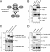
Interaction of pUL69 with Pin1. (A) Amino acid alignment of one of the tandem heptarepeats of the RNA-Pol II CTD and pUL69aa45-51. Phosphorylation sites for cellular CDK7 or CDK9, or viral pUL97 are highlighted. The Pin1 interaction motif within the RNA-Pol II CTD is boxed. (B, C, and D) CoIP experiments with HEK293T cells transfected with plasmids encoding the indicated proteins. As a negative control, either an empty vector (B, lanes 1 and 3, and C, lane 1) or an expression plasmid for FLAG-UAP56 (D, lane 1) was cotransfected. Two days later, the cells were lysed, and immunoprecipitation was performed using either anti-HA antibody (B) or polyclonal anti-Pin1 antibody (C and D). The coprecipitated proteins were stained by an anti-FLAG antibody.
FIG 5
FIG 5
Phosphorylation at S/T residue 46, 49, 51, or 52 does not affect the secondary structure of pUL69. (A) Schematic representation of the pUL69 N terminus with functional characteristics as described in the legend to Fig. 1. Confirmed phosphorylation sites are indicated by a “P” above the pUL69 amino acid sequence of HCMV strain AD169. (B) Amino acid sequences of wild-type pUL69aa47-77 (peptide 3) and its phosphorylated derivative pUL69aa47-77-pS51,pT52 (peptide 4); wild-type peptide 5, comprising amino acids 38 to 67; or its derivative with pS49 (peptide 6) or double-phosphorylated pS46 plus pS49 (peptide 7). (C) Empirical structure prediction of peptide 3 as determined by the Phyre2 server (see the text for details). (D and E) Schematic representations of the secondary structures of both peptides as determined by the chemical shift differences (in parts per million) of the α-protons between the experimental values and those for residues in a random coil for the indicated N-terminal pUL69 peptides obtained at 300 K in 50% aqueous TFE at pH 3. The localizations of the phosphorylation sites are indicated by arrows.
FIG 6
FIG 6
Pin1-induced cis/trans isomerization (c/t) of pUL69. (A to F) NMR spectroscopy of pUL69 peptides 6 (containing pS49) (A and D) and 7 (containing pS46 and pS49) (B, C, E, and F) in the presence or absence of Pin1 or Pin1 and the Pin1 inhibitor juglone. Superimposed expanded HN-HN regions (A to C) and HB regions (D to F) of the 2D 1H-1H NOESY (A to D) and 2D 1H-1H ROESY (E to F) spectra are depicted for phosphorylated (peptides 6 and 7) versions of a pUL69 peptide comprising amino acids 38 to 67. Phosphorylated peptides prior to (red signals) and after (blue signals) addition of Pin1 are shown; the green signals show the spectra obtained in the presence of Pin1 and juglone. Note the appearance of exchange peaks originating from an enhanced prolyl cis/trans interconversion rate after addition of Pin1 and that no exchange peaks are observed after addition of the Pin1 inhibitor juglone.
FIG 7
FIG 7
Characterization of recombinant cytomegaloviruses carrying amino acid substitutions for phosphorylation sites within the pUL69 N terminus. (A and B). Multistep growth curve analyses of HFFs that were infected with equal numbers of infectious units (MOI) of wild-type HCMV AD169, the UL69 deletion virus AD169-ΔUL69, or the mutant viruses AD169-UL69S/T1 and -S/T2. Viral supernatants were harvested at the indicated time points (dpi, days postinfection), followed by the determination of viral genomes released into the supernatants by quantitative real-time PCR. Each infection was performed in triplicate, and the standard deviations are shown.
FIG 8
FIG 8
Mapping of the UAP56/URH49 interaction motif in pUL69. Shown are CoIP analyses using cell lysates of HEK293T cells that were cotransfected with expression constructs for Myc-URH49, together with constructs for pUL69. (A and B) Plasmids expressing truncated pUL69 fused to FLAG-NLS-GST were used for cotransfection. (C and D) Plasmids expressing the N-terminal 146 amino acids of pUL69 with internal deletions or point mutations, as indicated, were used for cotransfection. Two days posttransfection, the cells were lysed, and immunoprecipitation was performed using anti-Myc antibodies. After electrophoresis, the coprecipitated proteins were visualized by Western blotting using anti-FLAG antibody (bottom [CoIP]). (C) Arginines required for UAP56 recruitment are indicated by asterisks; short linear motifs similar to the UBM are underlined.
FIG 9
FIG 9
UAP56/URH49 interaction of cellular and viral mRNA export factors. (A) UBMs comprising the short linear sequence SLDD or LDXXLD (indicated by solid underlining) found in the cellular mRNA export factors REF, UIF, CHTOP, LUTZP4, and SKAR. Positively charged residues are indicated in red, and negatively charged residues are in blue. Serines and leucines are highlighted in boldface. (B) UAP56/URH49 binding motifs of the betaherpesviral mRNA export factors UL69, C69, and Rh69. Amino acid stretches with similarity to the SLDD motif are underlined with dashed lines. Phosphorylation sites identified by MS analysis are circled and labeled as a solid P. Putative phosphorylation sites are circled and labeled as an open P. Residues crucial for UAP56/URH49 interaction that were identified in a previous study are indicated by open arrows. Serines 13 and 15, which were identified in this study as being critical for UAP56/URH49 interaction, are indicted by filled arrows. The putative UAP56 interaction regions of viral and cellular mRNA export factors are boxed.

References

    1. Sandri-Goldin RM. 2001. Nuclear export of herpes virus RNA. Curr Top Microbiol Immunol 259:2–23. - PubMed
    1. Sandri-Goldin RM. 2008. The many roles of the regulatory protein ICP27 during herpes simplex virus infection. Front Biosci 13:5241–5256. doi:10.2741/3078. - DOI - PubMed
    1. Toth Z, Stamminger T. 2008. The human cytomegalovirus regulatory protein UL69 and its effect on mRNA export. Front Biosci 13:2939–2949. doi:10.2741/2899. - DOI - PubMed
    1. Winkler M, Stamminger T. 1996. A specific subform of the human cytomegalovirus transactivator protein pUL69 is contained within the tegument of virus particles. J Virol 70:8984–8987. doi:10.1128/JVI.70.12.8984-8987.1996. - DOI - PMC - PubMed
    1. Winkler M, Rice SA, Stamminger T. 1994. UL69 of human cytomegalovirus, an open reading frame with homology to ICP27 of herpes simplex virus, encodes a transactivator of gene expression. J Virol 68:3943–3954. doi:10.1128/JVI.68.6.3943-3954.1994. - DOI - PMC - PubMed

Publication types