

Candida auris Forms High-Burden Biofilms in Skin Niche Conditions and on Porcine Skin
- PMID: 31969479
- PMCID: PMC6977180
- DOI: 10.1128/mSphere.00910-19
Candida auris Forms High-Burden Biofilms in Skin Niche Conditions and on Porcine Skin
Abstract
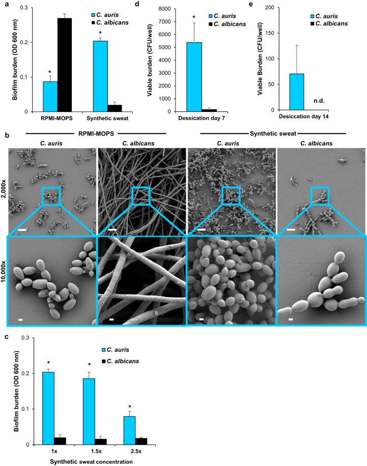
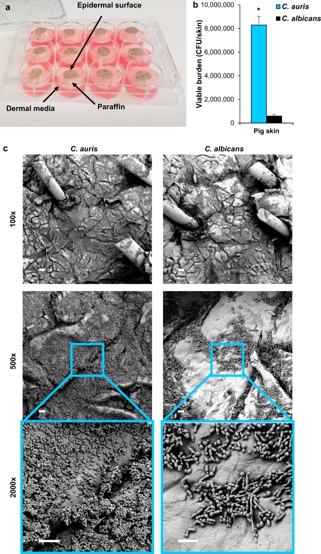
Emerging pathogen Candida auris causes nosocomial outbreaks of life-threatening invasive candidiasis. It is unclear how this species colonizes skin and spreads in health care facilities. Here, we analyzed C. auris growth in synthetic sweat medium designed to mimic axillary skin conditions. We show that C. auris demonstrates a high capacity for biofilm formation in this milieu, well beyond that observed for the most commonly isolated Candida sp., Candida albicans The C. auris biofilms persist in environmental conditions expected in the hospital setting. To model C. auris skin colonization, we designed an ex vivo porcine skin model. We show that C. auris proliferates on porcine skin in multilayer biofilms. This capacity to thrive in skin niche conditions helps explain the propensity of C. auris to colonize skin, persist on medical devices, and rapidly spread in hospitals. These studies provide clinically relevant tools to further characterize this important growth modality.IMPORTANCE The emerging fungal pathogen Candida auris causes invasive infections and is spreading in hospitals worldwide. Why this species exhibits the capacity to transfer efficiently among patients is unknown. Our findings reveal that C. auris forms high-burden biofilms in conditions mimicking sweat on the skin surface. These adherent biofilm communities persist in environmental conditions expected in the hospital setting. Using a pig skin model, we show that C. auris also forms high-burden biofilm structures on the skin surface. Identification of this mode of growth sheds light on how this recently described pathogen persists in hospital settings and spreads among patients.
Keywords: Candida auris; biofilm; pathogenicity; porcine; skin; sweat; transmission.
Copyright © 2020 Horton et al.
Figures
Comment in
- https://doi.org/10.1128/mSphere.00972-19 doi: 10.1128/mSphere.00972-19
References
-
- Lockhart SR, Etienne KA, Vallabhaneni S, Farooqi J, Chowdhary A, Govender NP, Colombo AL, Calvo B, Cuomo CA, Desjardins CA, Berkow EL, Castanheira M, Magobo RE, Jabeen K, Asghar RJ, Meis JF, Jackson B, Chiller T, Litvintseva AP. 2017. Simultaneous emergence of multidrug-resistant Candida auris on 3 continents confirmed by whole-genome sequencing and epidemiological analyses. Clin Infect Dis 64:134–140. doi: 10.1093/cid/ciw691. - DOI - PMC - PubMed
-
- Rudramurthy SM, Chakrabarti A, Paul RA, Sood P, Kaur H, Capoor MR, Kindo AJ, Marak RSK, Arora A, Sardana R, Das S, Chhina D, Patel A, Xess I, Tarai B, Singh P, Ghosh A. 2017. Candida auris candidaemia in Indian ICUs: analysis of risk factors. J Antimicrob Chemother 72:1794–1801. doi: 10.1093/jac/dkx034. - DOI - PubMed
-
- Schelenz S, Hagen F, Rhodes JL, Abdolrasouli A, Chowdhary A, Hall A, Ryan L, Shackleton J, Trimlett R, Meis JF, Armstrong-James D, Fisher MC. 2016. First hospital outbreak of the globally emerging Candida auris in a European hospital. Antimicrob Resist Infect Control 5:35. doi: 10.1186/s13756-016-0132-5. - DOI - PMC - PubMed
-
- Adams E, Quinn M, Tsay S, Poirot E, Chaturvedi S, Southwick K, Greenko J, Fernandez R, Kallen A, Vallabhaneni S, Haley V, Hutton B, Blog D, Lutterloh E, Zucker H, Candida auris Investigation Workgroup. 2018. Candida auris in healthcare facilities, New York, USA, 2013–2017. Emerg Infect Dis 24:1816–1824. doi: 10.3201/eid2410.180649. - DOI - PMC - PubMed
Publication types
MeSH terms
Grants and funding
LinkOut - more resources
Full Text Sources
Medical
