. 2020 Jan 22;10(1):924.
doi: 10.1038/s41598-020-57607-5.

Clinical and serological evaluation of capybaras (Hydrochoerus hydrochaeris) successively exposed to an Amblyomma sculptum-derived strain of Rickettsia rickettsii

Affiliations

Clinical and serological evaluation of capybaras (Hydrochoerus hydrochaeris) successively exposed to an Amblyomma sculptum-derived strain of Rickettsia rickettsii

Alejandro Ramírez-Hernández et al. Sci Rep. .

Erratum in

Abstract

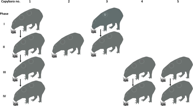
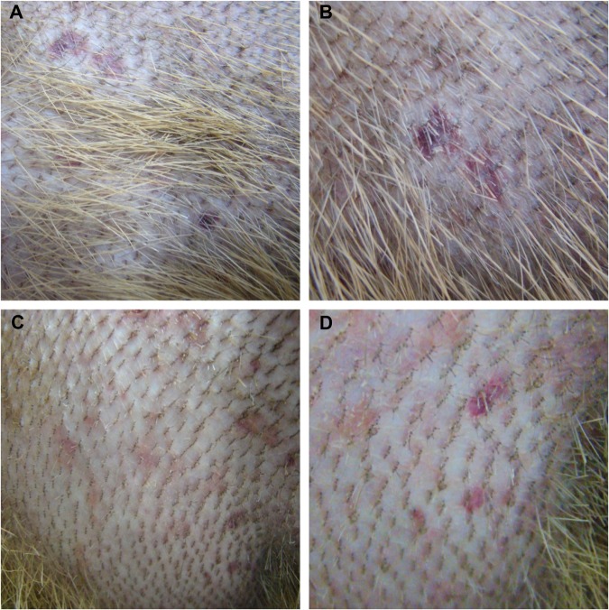
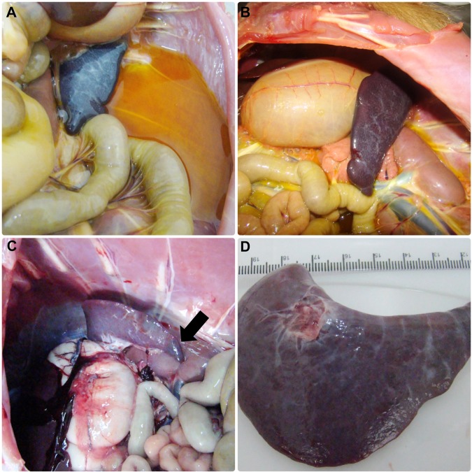
Brazilian spotted fever (BSF), caused by Rickettsia rickettsii, is the most lethal tick-borne disease in the western hemisphere. In Brazil, Amblyomma sculptum ticks are the main vector. Capybaras (Hydrochoerus hydrochaeris), the largest living rodents of the world (adults weighing up to 100 Kg), have been recognized as amplifying hosts of R. rickettsii for A. sculptum in BSF-endemic areas; i.e., once primarily infected, capybaras develop bacteremia for a few days, when feeding ticks acquire rickettsial infection. We conducted experimental infections of five capybaras with an A. sculptum-derived strain of R. rickettsii and performed clinical and bacteremia evaluation during primary and subsequent infections. Bacteremia was detected in all capybaras during primary infection, but not in subsequent infections. All animals seroconverted to R. rickettsii (titres range: 64-32,768), and remained seropositive throughout the study. Primary infection resulted in clinical spotted fever illness in four capybaras, of which two had a fatal outcome. Subsequent infections in seropositive capybaras resulted in no clinical signs. Capybaras developed a sustained immune response that prevented a second bacteremia. This condition may imply a high reproduction rate of capybaras in BSF-endemic areas, in order to continuously generate capybaras susceptible to bacteremia during primary infection.


Conflict of interest statement

The authors declare no competing interests.

Figures

Figure 1
Figure 1
Scheme of the experimental infections conducted on capybaras no. 1 to 5 during the study. Phase I, primary infection of capybara no. 1, and retaining capybara no. 3 as noninfected control; phase II, second infection of capybara no. 1 and primary infection of capybaras no. 2 and 3; phase III, third infection of capybara no. 1 and primary infection of capybaras no. 4 and 5; phase IV, fourth infection of capybara no. 1 and second infection of capybaras no. 4 and 5.
Figure 2
Figure 2
Skin of capybaras no. 2 and 3 during primary infection (phase II) with Rickettsia rickettsii (strain Itu) via tick exposure. (A,B) Purplish macules in capybara no. 2 (18 DPI). (C,D) Abdominal rash in capybara no. 3 (10 DPI). This figure has been published within the Doctoral Thesis of the first author (A. Ramírez-Hernández), which is available at the University of São Paulo’s digital library of Theses and Dissertations: https://teses.usp.br/teses/disponiveis/10/10134/tde-09092019-112817/en.php.
Figure 3
Figure 3
Abdominal cavity and spleen of capybaras no. 2 and 3 after primary infection (phase II) with Rickettsia rickettsii (strain Itu) via tick exposure. (A) Abdominal cavity of capybara no. 3 with evidence of ascites, jaundice and spleen enlargement (16 DPI) (B) Spleen enlargement in capybara no. 3 (16 DPI). (C) Spleen enlargement with apical hemorrhage (arrow) in capybara no. 2 (18 DPI) (D) Enlarged spleen from capybara no. 2 (18 DPI). This figure has been published within the Doctoral Thesis of the first author (A. Ramírez-Hernández), which is available at the University of São Paulo’s digital library of Theses and Dissertations: https://teses.usp.br/teses/disponiveis/10/10134/tde-09092019-112817/en.php.
Figure 4
Figure 4
Lung and stomach of capybara no. 2 after infection (phase II) with Rickettsia rickettsii (strain Itu) via tick exposure. (A) Lung of capybara no. 2 with evidence of bilateral disseminated vascular injuries (18 DPI). (B) Stomach of capybara no. 2 with an extended area of hemorrhage in the mucosa (18 DPI). This figure has been published within the Doctoral Thesis of the first author (A. Ramírez-Hernández), which is available at the University of São Paulo’s digital library of Theses and Dissertations: https://teses.usp.br/teses/disponiveis/10/10134/tde-09092019-112817/en.php.
Figure 5
Figure 5
Histopathological and immunohistochemical evaluation in an experimental infection of Capybaras (Hydrochoerus hydrochaeris) with Rickettsia rickettsii. (A) Inflammation and vasculitis in heart (arrow) (hematoxylin and eosin staining, objective 4x). (B) Inflammatory cell infiltrate in kidney (arrow) (hematoxylin and eosin staining objective 4x). (C) Liver with vasculitis and microthrombi (arrow) (hematoxylin and eosin staining objective 10x). (D) Brain; immunostaining of Rickettsia rickettsii in vessels (red bacili) (arrow), immunoalkaline phosphatase staining, naphthol fast red substrate with hematoxylin counterstain (objective 100x).
Figure 6
Figure 6
Rickettsia rickettsii antibody titres (IFA) after multiple infections with R. rickettsii (strain ITU) via tick exposures, in capybaras no. 1, 4 and 5. Dashed arrows indicate 2nd, 3rd and 4th infection of capybara no. 1 at 120, 248 and 475 days post primary infection (DPI), respectively. Straight arrow indicates 2nd infection of capybaras no. 4 and 5, 227 DPI. This figure has been published within the Doctoral Thesis of the first author (A. Ramírez-Hernández), which is available at the University of São Paulo’s digital library of Theses and Dissertations: https://teses.usp.br/teses/disponiveis/10/10134/tde-09092019-112817/en.php.
Figure 7
Figure 7
Hematological variables evaluated in capybaras (Hydrochoerus hydrochaeris) according to days post primary infection (DPI) with Rickettsia rickettsii (strain Itu) via tick exposure. Capybara numbers 1 (X), 3 (control) (△), 2 (+), 3 (○), 4 (◇) and 5 (□). This figure has been published within the Doctoral Thesis of the first author (A. Ramírez-Hernández), which is available at the University of São Paulo’s digital library of Theses and Dissertations: https://teses.usp.br/teses/disponiveis/10/10134/tde-09092019-112817/en.php.
Figure 8
Figure 8
Molecular detection of Rickettsia rickettsii DNA in blood (X) and skin (○) samples in capybaras (Hydrochoerus hydrochaeris) according to days post primary infection (DPI) with R. rickettsii (strain Itu) via tick exposure.

References

    1. Oliveira SV, et al. An update on the epidemiological situation of spotted fever in Brazil. J. Venom. Anim. Toxins Incl. Trop. Dis. 2016;22:22. doi: 10.1186/s40409-016-0077-4. - DOI - PMC - PubMed
    1. Parola P, et al. Update on tick-borne rickettsioses around the world: a geographic approach. Clin. Microbiol. Rev. 2013;26:657–702. doi: 10.1128/CMR.00032-13. - DOI - PMC - PubMed
    1. Labruna MB. Ecology of Rickettsia in South America. Ann. N. Y. Acad. Sci. 2009;1166:156–166. doi: 10.1111/j.1749-6632.2009.04516.x. - DOI - PubMed
    1. Szabo MP, Pinter A, Labruna MB. Ecology, biology and distribution of spotted-fever tick vectors in Brazil. Front. Cell. Infect. Microbiol. 2013;3:27. doi: 10.3389/fcimb.2013.00027. - DOI - PMC - PubMed
    1. Burgdorfer W, Brinton LP. Mechanisms of transovarial infection of spotted fever Rickettsiae in ticks. Ann. N. Y. Acad. Sci. 1975;266:61–72. doi: 10.1111/j.1749-6632.1975.tb35088.x. - DOI - PubMed

Publication types